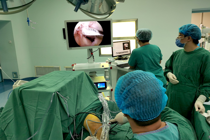
【Orthopedic Arthroscopy】Arthroscopic osteochondral autotransplantation
Release time: 08 Jul 2025 Author:Shrek
Arthroscopic osteochondral autografting is one of many methods for treating cartilage injuries. Osteochondral grafting is the transplantation of a healthy plug containing articular cartilage, cartilage ridges, and subchondral bone into an area that matches the size of the injury. Advantages of this technique include the use of articular hyaline cartilage rather than fibrocartilage to repair the defect and maintain joint height and shape. Arthroscopic osteochondral autotransplantation is a one-stage operation, is low-cost, and can even be completed in an outpatient setting. Full-scope surgery is technically challenging. Due to limitations in material collection, this technique cannot completely treat large-scale cartilage defects.

Indications and contraindications for surgery
Indications for cartilage autotransplantation include independent, full-thickness cartilage defects with a diameter of 1 to 2.5 cm. Large defects (diameter greater than 2.5cm) are less effective. In addition, this technique is generally limited to cartilage lesions with subchondral bone loss no deeper than 6 mm. Autologous cartilage transplantation is also not suitable for knees with damage to adjacent articular cartilage (equivalent to Type IV damage to tibial cartilage), multiple Type IV cartilage injuries, and knees that are unstable or poorly aligned. If the patient is older than 35 years old, the expected effect will be discounted.Some authors believe that patients older than 50 years should not use this technique. Other contraindications include a history of knee infection, intra-articular fracture, rheumatoid arthritis, and widespread degenerative arthritis. Meniscal tears and ligamentous instability are not absolute contraindications, but such conditions must be managed during cartilage transplantation. Autologous cartilage grafting is most commonly used for the femoral condyle, although autografting has been reported for tibial plateau, trochlear, and patellar lesions.
The Osteochondral Transplantation (COR) system can accurately obtain osteochondral plugs and implant them into drill holes in the defect area of the same size. The distinguishing features of the COR system are the cutting teeth of the extraction casing for more precise cutting depth, as well as the carefully designed drill bit for defect area preparation. This drill bit makes it easier to make the recipient hole perpendicular to the adjacent articular cartilage surface, making the size of the recipient and donor sites more consistent.
Surgical technique
Begin by performing a thorough exploratory arthroscopic assessment of the knee. When a localized, full-thickness cartilage defect is discovered, it is important to explore all areas of the knee joint, including the posterior recess and inferior meniscus, to identify and remove any mobile cartilage fragments. Arthroscopic grafting is suitable for most defect lesions; however, large and more posterior defects require extreme knee flexion to achieve an angle perpendicular to the articular cartilage, and sometimes a limited joint incision is required to achieve this angle. Use a lumbar needle to determine the optimal angle of entry, ensuring that the approach is perpendicular to the recipient and donor sites.
Arthroscopic autologous cartilage transplantation is completed in four steps:
1. Assess and prepare the defect area;
2. Determine the number of grafts;
3. Obtain materials;
4. Prepare the implantation area and implant the autologous plug.
Assessment and preparation of defective area
The knee joint and defect area should be carefully evaluated to ensure that the selection criteria are met and that no contraindications to this procedure exist. Defect preparation involves removing all free articular cartilage debris and using a curette or arthroscopic knife to create a vertical cartilage wall at the edge of the defect. The residual articular cartilage on the subchondral bone surface is removed, but extensive bleeding on the bone surface should be avoided. For better planning of bone grafting, the initially implanted plug should be placed in the most anterior part of the defect area immediately adjacent to the articular cartilage.

Determine the amount of bone to implant
Once the defect boundary is determined, a probe can be used to estimate the number of bone grafts required, or a sampling cannula can be used to measure the size and depth of the defect to determine which shape of post is most suitable for the defect area. The depth of the defect can be estimated using the COR system, which uses a probe or measuring tape on the side of the extraction casing. Generally, a series of 6 mm diameter graft plugs can be arthroscopically implanted and filled into the defect area. Larger posts are available, but they often require insertion through small incisions and tend to involve cartilage in high-load-bearing areas of the donor site.
The depth of the defect area should also be analyzed. Most defects have no obvious bone loss. In these cases, the standard 8 mm harvesting cannula is deep enough to fill the defect area. However, some defects (especially those with chondritis dissecans) have significant bone loss and must be addressed. In this case, bone grafting can be performed in the bone defect area in one operation, and cartilage transplantation can be performed later, or longer plugs can be obtained using harvesting cannulas of different depths, and the grafts can be placed in place so that the cartilage surface is flush with the surrounding cartilage surface, but the cancellous bone under the plug cartilage will be exposed at the base of the bone depression.
In transplantation, it is also important to evaluate the shape of the donor and recipient articular cartilage to achieve the best possible match of the cartilage surfaces. For large defects, the use of multiple posts can effectively reconstruct the original shape of the condyle. Although the use of smaller posts allows for better reshaping of the contour, the benefits are offset by reduced strength and stability of the graft and additional surgical steps are required.
- Recommended news
- 【General Surgery Laparoscopy】Cholecystectomy
- Surgery Steps of Hysteroscopy for Intrauterine Adhesion
- [Otolaryngology Nasal Endoscopy] How to Treat Recurrent Rhinitis
- [ENT Surgery: Nasal Endoscopy] Endoscopic Treatment of Nasal Polyps
- [Otolaryngology Nasal Endoscopy] Methods and Precautions for Nasal Bleeding Control under Nasal Endoscopy