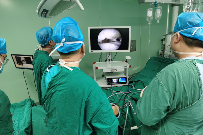
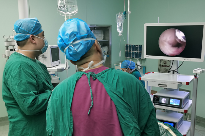
[Orthopedic Arthroscopy] Arthroscopic meniscectomy
Release time: 01 Jul 2025 Author:Shrek
Surgical principles
At present, the overall treatment principle for meniscal tears under arthroscopy is to preserve the meniscal tissue as much as possible, and the possibility of suturing and fixation should be considered before resection. For meniscectomy, the resection principle is to be as conservative as possible, only resecting the diseased part of the meniscus, and retaining as much healthy meniscal tissue as possible. The indication for meniscectomy is a clinically clearly diagnosed meniscal tear in patients who are not suitable for meniscal suturing and freshening. It is generally believed that meniscal injuries are located in areas without blood supply and have no healing potential; large compound tears, degenerative tears, flap tears, and most radial tears; those with difficulty in fixation and healing; older patients (45 to 50 years old) should all undergo meniscectomy.

Arthroscopic meniscectomy has different descriptions depending on the degree of meniscal tissue removed. Complete meniscectomy refers to the removal of all meniscal fibrocartilaginous tissue; subtotal meniscectomy refers to the removal of more than 50% of the meniscus tissue, but retains the rest of the meniscus. Peripheral annulus fibrosus; partial meniscectomy means that less than 50% of the meniscus tissue is removed and the annulus fibrosus is retained; the annulus fibrosus of the meniscus is transected during the operation, although some meniscal material is still retained, which can be called a functional meniscectomy because the meniscus has lost its function at this time.
Principles of arthroscopic meniscectomy:
①The scope of resection should be as conservative as possible;
②The attachment part of the meniscal joint capsule, that is, the annulus fibrosus, should be preserved;
③ Meniscal fragments with abnormal activity should be removed;
④ The meniscus edge shape should maintain gradual changes to avoid "sudden" changes and prevent mechanical symptoms after surgery.
Therefore, during meniscectomy, it is still necessary to follow the principles proposed by Metcalf et al. and pay attention to the details of the operation, in order to ultimately achieve good results.
Arthroscopic portal
Basic entrance:
① Upper inner side (SM): used to place the water inlet casing.
② Anterolateral (AL): used for placement of arthroscope.
③ Anteromedial (AM): used to place instruments (such as planers, basket forceps).
Surgical technique
The surgery adopts a conventional approach, but appropriate adjustments can be made to facilitate the operation. When establishing the second entrance, use a lumbar puncture needle to test the meniscus injury site and observe whether the instrument is easy to reach. For tears of the posterior horn of the medial meniscus, since the medial femoral condyle is larger and the curvature of the medial tibial plateau is deeper, the medial approach should be set lower to make it easier for instruments to treat the lesion, while the lateral approach should be set higher to enable more comprehensive arthroscopic observation. During the operation, it is necessary to exchange the arthroscope and instrument approaches at any time, observe the meniscus injury site from different angles, and use instruments to treat the diseased site.
During the operation, probes need to be repeatedly used for exploration, combined with the tear site diagnosed before surgery, and a combination of comprehensive and key examinations to avoid missing lesions. Lesions in the posterior horn of the meniscus are easy to miss and difficult to operate, so they require special attention. For patients with tight joints, comprehensive arthroscopic examination and treatment of the posterior horn of the medial meniscus is particularly difficult. In fact, the key lies in exposure. For medial meniscal injuries, it is recommended to add a baffle on the outside of the operating table and place it at the proximal femur and the balloon tourniquet. Applying valgus stress at the time of surgery provides a fulcrum to help open and expose the posterior aspect of the medial joint space. For lateral meniscus tears, place the knee joint in the figure-4 position, place the foot on the opposite calf, and apply downward pressure on the knee to open the lateral joint space.
Goals of arthroscopic meniscectomy:
①Remove unstable tear flap;
②The resection edge is trimmed into an arc shape;
③ Preserving the integrity and width of the meniscus ring as much as possible is of great significance for preserving function;
④Prevent damage to surrounding cartilage.
In order to achieve the above goals, during the resection process, it is necessary to repeatedly use probes to check the resection range and the stability of the residual meniscus to prevent residual lesions. According to literature reports, failure to detect and treat tears in other parts is an important reason for surgical failure. At the same time, avoid removing too much healthy meniscal tissue. Normal meniscal tissue has a tough feel during resection, so pay attention to identification. The meniscal tissue can be gradually removed and probed at any time to ensure that stable meniscal tissue is retained. From a biomechanical point of view, the annulus fibrosus at the edge of the meniscus plays an important biomechanical function, so it is very important to leave a continuous annulus fibrosus edge.
Finally, the free edge of the meniscus needs to be trimmed with a planer into a slope shape similar to that of a normal meniscus. At the same time, there is a smooth transition surface between the meniscus resection site and the normal part to avoid residual mechanical symptoms after surgery. In recent years, newly introduced radio frequency plasma equipment has been introduced to help treat menisci. Depending on the settings, it can cut the meniscus and trim the surface of the meniscus, which is better than manual equipment and planing systems.
Diagnostic arthroscopy
Examine the intraarticular and lateral compartments to identify displaced meniscal tissue.
medial compartment
Observe the medial meniscus and probe sequentially starting from the posterior side and moving forward along the superior and inferior surfaces to identify any displacement between the meniscal body and the tibial plateau.
If a tear is found, explore to determine its stability.
If the medial meniscus can be pulled forward over the midline of the medial femoral condyle, the diagnosis of medial meniscal tear and instability is confirmed.
Applying external stress to the posteromedial aspect of the knee can help expose the posterior meniscal horn.
lateral compartment
The knee joint was flexed 20° and turned into varus and internal rotation.
If arthroscopy is performed with the lower extremity extended, place the knee joint in the "4" position to facilitate inspection of the lateral compartments.
Systematic evaluation should begin at the posterior horn of the lateral meniscus, including the meniscal root.
The midsection of the lateral meniscus around the popliteus hiatus is evaluated by moving the arthroscope laterally and superiorly and inferiorly.
posterolateral compartment
A 70° arthroscope was inserted through the AM portal, passed through the intercondylar fossa above the anterior cruciate ligament (ACL), and past the lateral femoral condyle to examine the posterolateral compartment.
Posteromedial compartment (PM compartment)
To visualize the PM compartment, a blunt trocar is inserted through the AL portal, posteriorly and inferiorly along the medial wall of the intercondylar notch below the posterior cruciate ligament.
Usually, an auxiliary PM portal is created percutaneously at the posteromedial corner of the knee joint, a No. 18 spinal puncture needle is inserted into the compartment 1 cm above the medial joint space, and the skin is incised longitudinally with a No. 11 scalpel behind the medial femoral condyle.
Care is taken to avoid the saphenous nerve and vein, which can occasionally be visualized during penetration. A blunt trocar is then passed through the incision and the joint capsule is pierced under direct vision.
Meniscectomy for general tears
The goal of a partial meniscectomy is to remove part of the meniscal tissue that is unstable.
If the tear is deemed irreparable, all unstable torn meniscal edges and free meniscal fragments should be removed.
Using a planer rather than a basket forceps allows for more effective removal of fragmented meniscal tissue.
Articular cartilage must be protected. For knees with tight joint space, use the following tools to treat the meniscus (especially the medial meniscus).
Postoperative treatment
Rehabilitation training after arthroscopic meniscectomy is very important. The recovery process mainly depends on the extent of meniscus removal and the condition of the cartilage. Quadriceps isometric contraction exercises can be started on the day of surgery. Since most patients have quadriceps atrophy before surgery, quadriceps exercises need to be emphasized after surgery. After gaining quadriceps control, you can begin weight-bearing, initially using assistive devices, and transitioning to full weight-bearing, which generally takes 4 weeks. Range of motion training begins on the second day after surgery and progresses to a 90° extension and flexion range of motion within a few days based on the patient's tolerance. Postoperative use of nonsteroidal anti-inflammatory drugs can help reduce pain and swelling. Pain and discomfort at the incision site can last up to 6 months.

- Recommended news
- 【General Surgery Laparoscopy】Cholecystectomy
- Surgery Steps of Hysteroscopy for Intrauterine Adhesion
- [Otolaryngology Nasal Endoscopy] How to Treat Recurrent Rhinitis
- [ENT Surgery: Nasal Endoscopy] Endoscopic Treatment of Nasal Polyps
- [Otolaryngology Nasal Endoscopy] Methods and Precautions for Nasal Bleeding Control under Nasal Endoscopy