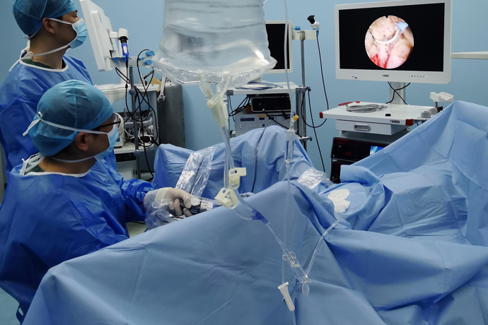
[Gynecological hysteroscopy] Hysteroscopic IUD removal
Release time: 27 May 2025 Author:Shrek
Everyone is familiar with the term "IUD". In medicine, we call it "intrauterine device".
In the 1970s and 1980s, family planning was listed as a basic national policy. Many people would use intrauterine devices to prevent pregnancy, which would not affect future pregnancy and could easily prevent pregnancy. According to statistics, from 1980 to 2009, Chinese women used intrauterine devices 286 million times. Thirty or forty years have passed, and the women who "put on the IUD" at that time have gradually entered menopause. Due to the neglect of IUDs and the lack of knowledge about IUDs, many women still do not realize that IUDs are not "once and for all" and need to be checked regularly to understand whether the IUD has shifted or fallen off. Otherwise, it will seriously endanger their health.

Hysteroscope is a fiber-light endoscope. Hysteroscopy refers to the use of uterine expansion medium to expand the uterine cavity, and inserting a light-guided glass fiber endoscope into the uterine cavity through the vagina to directly observe the physiological and pathological changes of the cervical canal, internal cervical os, uterine cavity and fallopian tube opening, so as to directly and accurately obtain samples of the diseased tissue and send them for pathological examination. At the same time, it can also be directly treated under hysteroscopy. It has the advantages of clear field of view, high lesion identification, small trauma, fast postoperative recovery, and day surgery. It is widely used in hospitals at all levels. Hysteroscopy and surgery have almost become a hurdle that female patients must face.
Hysteroscopy is used to treat difficult intrauterine devices (IUD). Most cases are caused by heavy menstruation or more than two failed attempts to remove the device during family planning. Hysteroscopy reveals that the IUD is twisted, deformed, or broken. Some IUDs are left with only remnants, some are implanted in the myometrium, and some are outside the uterine cavity. Hysteroscopy can directly observe the shape and position of the IUD in the uterine cavity, and whether it is implanted in the uterine wall, thereby determining the route and method of removing the IUD.
Common side effects or complications after IUD placement include:
1. Abnormal uterine bleeding and pain;
2. IUD incarceration and IUD ectopic placement;
3. Ectopic perforation;
4. Pregnancy with IUD (meaning pregnancy and IUD coexisting), etc.
Situations where intrauterine devices are not suitable:
1. Acute pelvic inflammatory disease; acute vaginitis;
2. Severe cervical erosion, menorrhagia or irregular bleeding, uterine fibroids, and narrow cervical opening;
3. Women with severe systemic diseases should not have the device placed, otherwise it will lead to aggravated inflammation and increased menstrual flow.
In what cases should the ring be removed:
1. Those who have had the IUD for more than 5-10 years and want to replace it with a new one.
2. Those who have irregular vaginal bleeding or other symptoms that are ineffective after treatment. Those with heavy vaginal bleeding or infection should remove the IUD in time.
3. Those who want to have another child.
4. Those who have been sterilized for 1 year. Those who have been menopausal for more than half a year are recommended to remove the IUD.
5. Those who have severe side effects and want to change their contraceptive method.
6. Those who are pregnant with the IUD can remove it during abortion.
IUD malposition means:
The IUD leaves its normal position in the uterine cavity and is partially or completely embedded in the myometrium, or is malpositioned in the abdominal cavity, broad ligament, bladder, rectum, etc. When the IUD is malpositioned, it not only no longer has a contraceptive effect, but also causes trouble to the user.
Symptoms of IUD misplacement:
1. Abdominal pain;
2. Pregnancy with IUD;
3. Continuous menstrual bleeding
Causes of IUD dislocation:
1. The risk of IUD dislocation increases when placed during lactation;
2. The IUD is too large or placed in the wrong position;
3. The shape of the IUD is not compatible with the shape of the uterine cavity
According to the degree of ectopicity, it can be divided into three categories:
(1) Partial incarceration of the IUD: The IUD is partially embedded in the endometrium and myometrium.
(2) Complete incarceration of the IUD: The IUD is completely embedded in the myometrium or mostly embedded in the myometrium with some of the uterine serosa exposed.
(3) Extrauterine ectopic IUD: The IUD is ectopically located in the pelvic cavity, abdominal cavity, bladder, intestine, broad ligament or outside the peritoneum.
Case
Ms. Chen had an intrauterine contraceptive ring inserted after giving birth to her second child 6 years ago. In the past two months, she often felt pain in her lower abdomen and occasionally had a small amount of vaginal bleeding. Ms. Chen rushed to the hospital for a color Doppler ultrasound examination, and the results showed that the intrauterine contraceptive ring was embedded in the muscle layer. In other words, the contraceptive ring was not working properly and had penetrated into the muscle layer of the uterus, requiring surgery.
The doctor quickly arranged a hysteroscopy. Under the microscope, it can be seen that the arms of the "V"-shaped IUD are deeply embedded in the uterine horn muscle layer. In this case, it is very dangerous to perform a normal IUD removal. If the IUD fails to be removed under blind exploration, the patient's pain will increase. Once the uterus is perforated, the consequences will be very serious. Therefore, the IUD was removed under hysteroscopy. The situation in the uterus is clear at a glance, and the IUD was successfully and completely removed. The patient does not need to be hospitalized or operated on. The process is basically painless. The operation time is only a few minutes, and the patient can leave the hospital immediately after the operation.
The doctor quickly arranged a hysteroscopy. Under the microscope, it can be seen that the arms of the "V"-shaped IUD are deeply embedded in the uterine horn muscle layer. In this case, it is very dangerous to perform a normal IUD removal. If the IUD fails to be removed under blind exploration, the patient's pain will increase. Once the uterus is perforated, the consequences will be very serious. Therefore, the IUD was removed under hysteroscopy. The situation in the uterus is clear at a glance, and the IUD was successfully and completely removed. The patient does not need to be hospitalized or operated on. The process is basically painless. The operation time is only a few minutes, and the patient can leave the hospital immediately after the operation.
Case
Aunt Liu, 60, has been in menopause for more than 10 years. When she was young, she had an IUD placed in her uterus for contraception. In the past half month, Aunt Liu has been feeling uncomfortable in her lower abdomen and wanted to take out the IUD. After examination, it was found that Aunt Liu's cervix had atrophied. In addition, she had undergone two cesarean sections, and her uterus and the anterior abdominal wall were densely adhered, resulting in the cervix being invisible. If you can't see the cervix, you can't take out the IUD at all! This is the same as not being able to find the door and not being able to enter the house at all. This worried Aunt Liu and her daughter.After discussion, the doctor decided that this problem could be solved through hysteroscopic surgery! Under direct vision of the hysteroscope, the doctor successfully found the cervical opening and entered the uterine cavity. It was found that Aunt Liu's IUD had been embedded in the flesh. With the help of hysteroscopy and ultrasound, it took only 10 minutes to successfully remove the IUD, solving the big problem that had troubled Aunt Liu.
In this operation, the natural vaginal cavity is used to achieve no incision and no scar. The intrauterine contraceptive ring is successfully removed by hysteroscopic endoscopy technology when the cervix is severely atrophied and cannot be exposed. It is really a "small hysteroscopy, solving a big problem"! Here, I would also like to remind all female friends that if you have been in menopause for half a year, please remove the intrauterine contraceptive device as soon as possible, because the uterus of women shrinks after menopause, but the size of the contraceptive ring remains unchanged, which greatly increases the risk of ectopic and incarcerated contraceptive rings!
Expert interpretation
In fact, situations like Ms. Chen and Aunt Liu's are not uncommon, and medically it is called IUD misplacement.
What are the causes of IUD dislocation?
1. Improper operation causes the IUD to be placed outside the uterine cavity;
2. The IUD is too large, too hard, or the uterine wall is thin and soft, and the uterine contraction causes the IUD to gradually dislocate outside the uterine cavity.
How to diagnose IUD malposition?
When the intrauterine contraceptive device is not in the normal position in the uterine cavity, it is called IUD malposition, including downward movement, incarceration, and external movement of the IUD.
1. The sonogram of the normal position of the metal IUD is a strong echo in the endometrial cavity with a low echo halo around the endometrium. In addition, the distance between the upper edge of the IUD and the uterine fundus or the distance between the lower edge of the IUD and the internal os of the uterus is measured to determine whether the IUD position is normal.
2. Grading of IUD downward movement Mild: The upper edge of the IUD is <25mm from the uterine fundus serosa; Moderate: The upper edge of the IUD is 25-35mm from the uterine fundus serosa; Severe: The upper edge of the IUD is >35mm from the uterine fundus serosa.
3. Grading of IUD embedded in the myometrium Mild: The depth of IUD embedded in the muscle wall is <1/3; Moderate: The depth of embedded in the muscle wall is 1/3~2/3; Severe: The depth of IUD embedded in the muscle wall is >2/3 or reaches the uterine serosa.
How to prevent IUD dislocation?
First, choose a regular medical institution to place an IUD. Second, complete the preoperative examination to rule out genital malformations (such as septate uterus, double uterus, etc.), genital tumors (such as submucosal fibroids) and serious systemic diseases. At the same time, choose a suitable type of IUD and place it within 3-7 days after the menstrual period ends.
What precautions should be taken after the placement of the IUD?
1. Avoid heavy physical labor within 1 week after placement.
2. Avoid bathing and sexual intercourse within 2 weeks.
3. Follow-up at 1, 3, 6, and 12 months in the first year after surgery, and follow-up once a year thereafter until discontinuation. See a doctor at any time for special circumstances.
4. Remove the IUD promptly when the placement period is reached.
Is it necessary to remove the IUD?
First of all, we should know that the IUD also has a time limit. If it exceeds its service life, it is recommended to remove it in time, whether considering the contraceptive effect or the occurrence of long-term complications.
For women within one year after menopause, there is no need for contraception, and the IUD needs to be removed in time. After menopause, the estrogen and progesterone levels in the body drop significantly, the uterus shrinks, the cervix becomes tighter, and the uterine muscle layer becomes thinner, while the size of the intrauterine contraceptive device remains unchanged. The shrunken uterus may be embedded in the uterine wall by the IUD, which is called IUD incarceration. If it is embedded in a blood vessel, it may cause heavy bleeding; if it wanders in the abdominal cavity, it may cause a series of abdominal complications (abdominal pain, infection, etc.); if it wanders to the bladder and is embedded in the bladder cavity, it may cause stones and cause unexplained hematuria (just like Ms. Wang's IUD malposition in the case). Any malposition can make it difficult or fail to remove the IUD, and even require laparotomy to remove the IUD.
Older women will inevitably get sick, and some diseases require MRI examinations, but women with IUDs generally cannot undergo MRIs. Therefore, for women who use intrauterine devices, once they are inserted, they need to be taken out later. Don't be lucky and think that since you don't feel anything, just leave it in the uterus. When you really feel something, I'm afraid you'll have to make a big move.
Therefore, I remind women of childbearing age and perimenopausal women again to choose a regular hospital to place the correct type of IUD. IUDs have a time limit. When they expire or there is no need for contraception, please remove the IUD in time.
Precautions before and after hysteroscopy
Before surgery:
1. It is best to perform hysteroscopy 3-7 days after menstruation.
2. Sexual intercourse is prohibited for three days after menstruation or before surgery.
3. Preoperative examinations include: leucorrhea routine, blood routine, coagulation function, four preoperative items, liver and kidney function, blood type identification, and electrocardiogram.
Postoperatively:
1. No sexual intercourse or bathing for 1 month after surgery;
2. Rest for at least 1 week after surgery;
3. Appropriate antibiotic treatment after surgery;
4. Come to the hospital for treatment at any time if there is excessive vaginal bleeding;
5. Come to the hospital for pathology report 7 days after surgery.

- Recommended news
- 【General Surgery Laparoscopy】Cholecystectomy
- Surgery Steps of Hysteroscopy for Intrauterine Adhesion
- [Otolaryngology Nasal Endoscopy] How to Treat Recurrent Rhinitis
- [ENT Surgery: Nasal Endoscopy] Endoscopic Treatment of Nasal Polyps
- [Otolaryngology Nasal Endoscopy] Methods and Precautions for Nasal Bleeding Control under Nasal Endoscopy