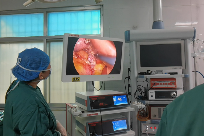
[Laparoscopic hepatobiliary surgery] fenestration and drainage of liver cysts
Release time: 10 Jun 2025 Author:Shrek
Laparoscopic liver cyst fenestration and drainage is used for the surgical treatment of liver cysts. Liver cysts are a common benign liver disease. When the liver cyst is larger than 10 cm or has compression symptoms, surgical treatment is required. Since the first report of laparoscopic liver cyst fenestration in 1991, it has been replacing open surgery due to its advantages of less trauma, faster recovery and better results.

Indications
Laparoscopic liver cyst fenestration and drainage is suitable for:
1. Single or single multi-chamber, symptomatic liver cysts, with shallow cysts and a thickness of no more than 1mm from the surface of liver tissue.
2. Liver cysts found during laparoscopic cholecystectomy.
Contraindications
1. Both communicating and tumorous liver cysts are contraindicated.
2. Multiple cysts and cysts located in the right posterior lobe.
3. Cysts in the liver parenchyma.
4. Patients with a history of upper abdominal surgery.
Preoperative preparation
1. The same as the routine preoperative preparation for laparotomy.
2. Liver B-ultrasound, CT or MRI examination is an indispensable and important examination, which can clarify the thickness of the liver tissue on the surface of the cyst, the relationship between the cyst and the intrahepatic blood vessels and bile ducts, and the surface location.
3. Functional measurement of important organs (heart, lung, liver, kidney, etc.).
4. Exclude hepatic echinococcosis and hepatic cystic tumors. If the cyst cannot be excluded from being connected to the bile duct, retrograde pancreaticobiliary angiography is required.
Detailed surgical steps
1. After anesthesia, disinfect and spread the towel, make a puncture hole under the navel, establish artificial pneumoperitoneum, and insert laparoscopic instruments according to the 3-hole method.
2. Exploration showed multiple cysts on the surface of the liver. A huge cyst with a diameter of about 20 cm was found in the right liver. Fibrosis was seen on the surface of the cyst wall and the blood supply was rich. The electrocoagulation hook was electrocuted at the thinner part of the cyst wall, and colorless and slightly turbid liquid was seen to spray out. The cyst fluid was drained with an aspirator, totaling about 2000 ml.
3. The ultrasonic knife was used to cut and stop bleeding along the edge of the junction between the cyst wall and the liver parenchyma. Obvious dilated blood vessels and flutter of the inferior vena cava were seen in the cyst cavity; another cyst was seen at the bottom of the cyst cavity, and the surface cyst wall was removed by ultrasonic knife to open the window and remove the cyst wall; multiple small cysts were seen in the liver parenchyma on the left side of the gallbladder, and part of the cyst wall was removed by ultrasonic knife to open the window, and the cyst wall was removed in batches.
4. Remove the cyst wall without liver parenchyma and put it into the specimen bag, and take it out through the umbilicus.
5. Use ultrasonic knife to take a section of greater omentum with vascular pedicle, fill it into the liver cyst cavity and fix it to the edge of the cyst wall with Hemolok clip.
6. Check that there is no active bleeding at the edge of the cyst, place the right subhepatic drainage tube in front of the right axilla, and place the liver cyst cavity drainage under the xiphoid process.
Postoperative care
1. Same as general liver surgery.
2. Encourage patients to get out of bed and move around as soon as possible to facilitate drainage and absorption of cystic fluid.
3. The drainage tube is connected to a sterile bag and removed within 2 to 3 days if no bile is drawn out.
4. B-ultrasound or CT scan should be repeated after surgery to understand the effect of the operation.
Precautions
After discharge, you should have a regular life, ensure adequate rest and sleep, and exercise regularly; quit smoking, drinking, coffee, strong tea, and carbonated drinks; chew food slowly and eat light and easy-to-digest food, and avoid spicy and greasy food; it is recommended to eat small meals frequently, and avoid excessive exercise after meals.
Postoperative diet
Usually, oral feeding can be started on the first day after surgery, starting with water, then gradually changing to liquid food, semi-liquid food, and finally normal food.

- Recommended news
- 【General Surgery Laparoscopy】Cholecystectomy
- Surgery Steps of Hysteroscopy for Intrauterine Adhesion
- [Otolaryngology Nasal Endoscopy] How to Treat Recurrent Rhinitis
- [ENT Surgery: Nasal Endoscopy] Endoscopic Treatment of Nasal Polyps
- [Otolaryngology Nasal Endoscopy] Methods and Precautions for Nasal Bleeding Control under Nasal Endoscopy