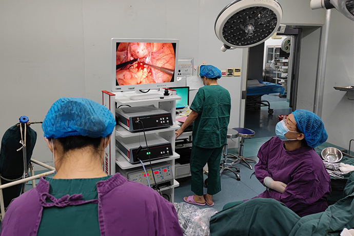
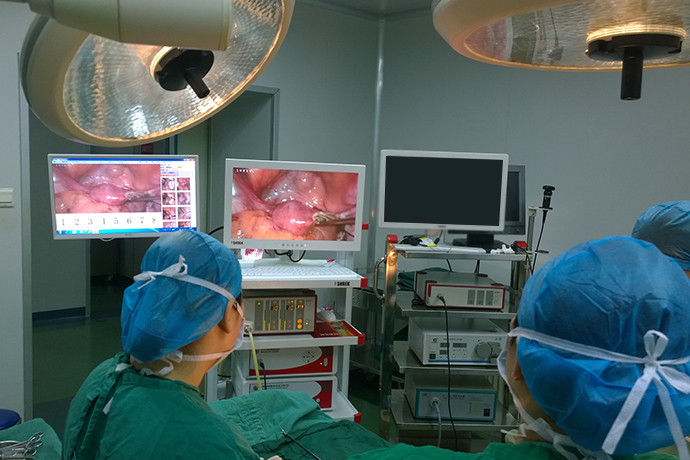
[Gynecological Laparoscopy] 4K Laparoscopy for Treatment of Hydrosalpinx
Release time: 16 Aug 2023 Author:Shrek
Why hydrosalpinx?
The fallopian tube is the pathway of sperm and the place where the sperm and egg combine. If the fallopian tube is narrowed, atresia, deformed or due to various reasons of adhesion, poor peristalsis caused by hormonal disorders, etc., sperm and eggs cannot meet or cannot be transported smoothly. Can cause infertility or ectopic pregnancy. Among them, hydrosalpinx is a typical pathological manifestation of inflammatory infection.

Why does hydrosalpinx occur?
The latest international statistics show that the incidence of tubal factor infertility accounts for about 30% to 40% of infertile women, suggesting that its occurrence is very common. Fallopian tube lesions are generally caused by pelvic inflammatory disease. These microbial pathogens enter the pelvic cavity through the female vagina and cause ascending infection. After reaching the fallopian tube cavity, they cause inflammatory changes, leading to tube wall congestion, suppuration, and exudation, and finally destroy the fallopian tube mucosa. It causes damage to endothelial cells, loss of ciliated cells, adhesions at the umbrella end, and accumulation of exudate and secretions in the lumen, resulting in expansion and thinning of the tube wall, thereby forming hydrosalpinx.
Hydrosalpinx is mainly related to the following factors
1. Pelvic cavity and genitourinary tract inflammation
2. Surgical injury
3. Postpartum and post-abortion infection
4. Genital tuberculosis
How is hydrosalpinx discovered?
The diagnosis of hydrosalpinx mainly relies on medical methods such as lipiodol radiography and pelvic ultrasound. Hysterosalpingography (HSG) can confirm the diagnosis. That is, a contrast agent is injected into the uterine cavity through the cervical canal, and the lumen is visualized under X-ray photography. However, if the angiography shows "water accumulation", but no signs of water accumulation are found in multiple ultrasound examinations, careful diagnosis is required, combined with medical history, pelvic bimanual examination, and comprehensive judgment to prevent overdiagnosis.
Treatment of hydrosalpinx
Was diagnosed with hydrosalpinx, the best solution is laparoscopic surgery.
Modern pathological studies have found that some unexplained hydrosalpinx may be the source and accompanying factors of ovarian serous cystadenocarcinoma. For the obvious hydrosalpinx, it tends to be removed.
The fallopian tube is not only a place for fertilization, but also has the function of transporting fertilized eggs. Therefore, a simple "salpinx dredging" operation cannot significantly improve the natural pregnancy rate of patients with hydrosalpinx. In addition, a number of international clinical studies have shown that resection of hydrosalpinx in patients with repeated V failures can significantly increase the pregnancy rate. Therefore, it is very important to accept the advice of professional doctors and perform reasonable tubal surgery according to the ovarian reserve function.
Can hydrosalpinx be treated laparoscopically?
Laparoscopic surgery is a minimally invasive diagnosis and treatment technique, and it is a treatment method recommended by many medical institutions. For different types of patients, according to the degree of hydrosalpinx, more appropriate treatment methods for hydrosalpinx can be selected. Laparoscopic surgery is the most effective method among the current treatment methods. It can treat other inflammations in the fallopian tube at the same time, and has a high medical effect. Laparoscopic treatment of hydrosalpinx mainly uses laparoscope and other instruments to carry out digital photography. Through this real-time whole-process monitoring, doctors analyze and judge the patient's condition, and use this judgment to carry out surgical treatment. The effect of this treatment is obvious, and the patient recovers quickly, and usually recanalizes at one time.
Advantages of Laparoscopy
1. Laparoscopy can be used to check from different angles and directions with an ultra-high-definition 4K monitor without affecting the abdominal organs, and even some deep positions can be seen, achieving the effect of visual inspection, without missed diagnosis or misdiagnosis.
2.4K laparoscopic surgery is carried out in a closed basin and abdominal cavity, with little interference from the internal environment. The trauma suffered by the patient is far less than that of the laparotomy, and the patient recovers quickly after the operation.
3. The operation is performed by a professional doctor, and the treatment can be completed in a short time without affecting normal physiological functions, and normal work and life can be resumed after the operation.
4.4K laparoscopic surgery has little disturbance to the internal organs of the abdominal cavity, and avoids the stimulation and pollution of the abdominal cavity by air and dust bacteria in the air. Ultrasonic scalpel and bipolar electrocoagulation are the main operations during the operation. The blood vessels are coagulated first and then cut off. The bleeding is completely stopped and the bleeding is very small. Before the operation is completed, the abdominal cavity is kept clean. Therefore, the postoperative intestinal function can be recovered quickly, and the food can be eaten earlier, which greatly reduces the factors of postoperative intestinal adhesion.
5.4K Laparoscopic surgery is the representative of truly minimally invasive surgery, the trauma is greatly reduced, the operation process and postoperative recovery are easy, and there is less pain.
6. You can get out of bed early after the operation, and the sleeping position is relatively random, which greatly reduces the intensity of accompanying nursing care for family members.
7. The holes in the abdominal wall are small (ranging from 3-10mm), scattered and concealed, and the appearance will not be affected after healing.
8. General anesthesia is generally used, and all monitoring is complete, which greatly increases the safety.
9. Poke hole infection is far less than traditional incision infection or fat liquefaction.
10. Abdominal wall poking holes replace the abdominal wall incision, avoiding the damage of abdominal wall muscles, blood vessels and corresponding nerves, and there will be no abdominal wall weakness and abdominal wall incisional hernia after surgery. Cutting causes numbness of the corresponding skin.
Case brief
Patient Zheng, female, 36 years old, has not been pregnant for 13 years without contraception, found a mass in the right adnexa for more than 2 years, and has fertility requirements.
Gynecological color Doppler ultrasound performed 2 years ago indicated that the mass in the right adnexal area (about 3*4cm) might be considered hydrosalpinx. A reexamination color Doppler ultrasound half a year ago indicated the possibility of a cystic mass (about 48*30mm in size) in the right adnexal area of the fallopian tube. Gynecological examination: palpable thickening in the right adnexal area, and no abnormalities in the rest.
Specific steps
1. Routine preoperative preparation, placement of urinary catheter and vaginal insertion of a radiography tube into the uterus. This contrast tube is used during the operation to check whether the fallopian tubes are unobstructed during the operation.
2. Cut through the layers of the abdominal wall according to the routine, and after exploring the pelvic cavity, lift the fallopian tubes to the operation field. Take the distal end of the fallopian tube and make a "ten" or "*" incision.
3. Inject physiological saline and methylene blue solution through the contrast tube to check whether the fallopian tube is unobstructed, and observe whether the mucosal folds in the lumen of the fallopian tube are abundant and whether the tissue is healthy.
4. The mucous membrane of the fallopian tube is turned out, and several short longitudinal incisions are made on the wall of the avascular area to make it into a petal shape or a natural umbrella shape.
5. Carefully suture the mucosal and serosal surfaces of the fallopian tube intermittently with 4-0 intestinal wound sutures, so that the incision edge of the fallopian tube has no bleeding and has a natural umbrella shape.
Postoperative care
(1) Use broad-spectrum antibiotics for at least 1 week. Antihistamine drugs were used as appropriate to reduce anastomotic edema.
(2) Postoperative fluid flow was performed 1 to 2 days earlier than anastomosis.
(3) If an indwelling stent is placed, it will be removed 10-14 days after the operation, and fluid will flow immediately after removal.

- Recommended news
- 【General Surgery Laparoscopy】Cholecystectomy
- Surgery Steps of Hysteroscopy for Intrauterine Adhesion
- [Otolaryngology Nasal Endoscopy] How to Treat Recurrent Rhinitis
- [ENT Surgery: Nasal Endoscopy] Endoscopic Treatment of Nasal Polyps
- [Otolaryngology Nasal Endoscopy] Methods and Precautions for Nasal Bleeding Control under Nasal Endoscopy