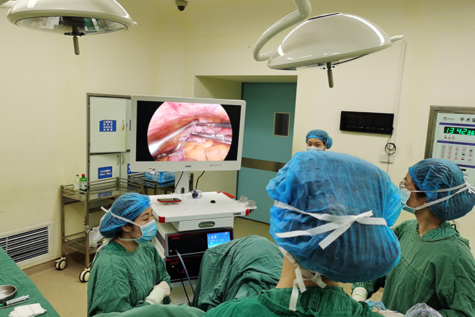
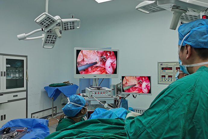
[Gynecological Laparoscopy] 4K Laparoscopic Subtotal Hysterectomy
Release time: 08 Aug 2023 Author:Shrek
Subtotal hysterectomy for women is to remove most of the uterine body and only retain the cervix. It is generally aimed at younger women suffering from adenomyosis or uterine fibroids. Surgical approach.
After subtotal hysterectomy, women will no longer have menstruation, and at the same time, normal pregnancy and childbirth will no longer occur.
After subtotal uterine surgery, the ovary can maintain normal endocrine function, and it does not affect women's sex life. However, because women have a cervix, as long as they have normal sex life, they need regular TCT examinations of the cervix to avoid Cervical chronic inflammation or cervical cancer.
In addition, women should also pay attention to keeping the vulva clean and hygienic to reduce the occurrence of vaginal bacterial ascending infection.

4K Full HD Laparoscopic Subtotal Hysterectomy (LSH):
LSH refers to laparoscopic removal of the uterus while preserving the cervix. It has the advantages of short operation time, less intraoperative bleeding, low postoperative morbidity, and quick recovery. It is a surgical procedure with practical significance in clinical practice.
Precautions for 4K laparoscopic subtotal hysterectomy During the operation, care must be taken not to damage the ureter and reduce blood loss as much as possible. For this reason, the operator must be familiar with the local anatomy of the uterus, especially the distribution of blood vessels and the location and direction of the ureter.
Surgical steps
1. In supine position or lithotomy position, place the palace lifter.
2. Treatment above the uterine vessels is the same as laparoscopic total hysterectomy.
3. After resection of the uterine blood vessels, 3 channels of the cervix were ligated at 1 cm below the planned resection plane of the isthmus, and cut off with electrocoagulation scissors above it. The mucous membrane of the tube should be electrocautery as far as possible, and care should be taken not to scald the tube.
4. Rinse the pelvis and stop the bleeding.
5. The bladder peritoneum was reflexed to cover the cervical stump, and several stitches were sutured to fix it. To prevent cervical prolapse, the ends of the round ligament can be sutured to the cervix.
6. Take out the uterine body Put it into the large claw forceps through the 10mm sheath, use a cylindrical rotary cutter to crush the tissue, and crush the uterine body into strips and take it out. It is more ideal to use electric tissue morcellator. If there is no morcellator, a small incision can be made on the original puncture hole of the abdominal wall, and the uterus can be taken out, or can be taken out through the posterior fornix incision.
7. Wash the abdominal cavity again, suck out the effusion, exhaust, take the trocar sheath and lens, and suture each puncture hole.
Precautions
1. Treatment of accessories If the patient needs to keep the accessories, cut off the proper ligament of the ovary, fallopian tube, and round ligament; The pelvic infundibulum ligament contains ovarian blood vessels, which can be closed by electrocoagulation and then cut to stop bleeding, or the peritoneum at the mesentery of the ovary can be opened first, and the pelvic infundibulum ligament can be ligated and then cut. When dealing with the tissues of the uterine horns, special attention should be paid to the branches of the uterine artery to the ovaries and fallopian tubes and their accompanying veins. The vein is located in the subperitoneum, if you do not pay attention, it is easy to tear and cause bleeding. Once bleeding is more troublesome to stop the bleeding. Therefore, when cutting these structures, you can stay away from the uterine horn, so that it is easier to coagulate, close and stop bleeding.
2. Treatment of the broad ligament When separating the broad ligament, the anterior and posterior peritoneum can be cut together without separation, and the lower edge of the incision can reach the level of bladder peritoneal reflexion, and care should be taken not to injure the uterine blood vessels. The ureter does not need to be separated, and generally will not be damaged. The incision of the broad ligament should be away from the uterine wall to avoid touching the ascending branch of the uterine artery ascending along the lateral wall of the uterus. If the fibroid is located in the broad ligament, it is necessary to open the peritoneum of the anterior and posterior leaves of the broad ligament, push the peritoneum away against the surface of the fibroid, and free the fibroid, so that the ureter will be pushed to the side wall of the pelvis without damage.
3. Bladder peritoneal reflex For patients without cesarean section history, the anatomy of the peritoneal reflex remains unchanged, and the peritoneum can be cut directly and the bladder pushed down. The gap between the bladder and the cervix is very clear and easy to push down. Use the vault cup to prop up the entire vault, making pushing down the bladder very easy. Generally speaking, the two sides of the cervix do not need to be pushed too far apart, so as not to cause bleeding. If there is a history of cesarean section, scars are often formed at the bladder peritoneal reflexion, so care should be taken not to damage the bladder when separating.
4. Treatment of uterine blood vessels The treatment of uterine blood vessels is the difficulty of total hysterectomy. If the uterine blood vessels are not handled properly and cause bleeding, it will affect the operation and even lead to complications. The main point of the treatment of uterine blood vessels is to dissect the uterine blood vessels clearly, and then close to the uterine side to block them. The commonly used method is to cut off the blood vessel after electrocoagulation. Uterine vessels can also be ligated with sutures or blocked with vascular clips. The uterine artery can also be ligated and cut off at the point where the uterine artery branches off from the internal iliac artery. The main point of this method is to cut the peritoneum between the garden ligament and the fallopian tube, separate the loose connective tissue inside it, separate it downward along the surface of the ureter, and cut it in the ureteral tunnel. The uterine artery is identifiable at its entrance. The uterine artery is divided retrogradely to the branch of the internal iliac artery, and the uterine artery can be blocked or cut off by electrocoagulation. It can also be cut from the posterior lobe of the broad ligament and above the ureter, separated toward the pelvic wall to find and block the uterine artery.
5. Cutting of the uterosacral ligament and main ligament Although there are no large blood vessels in these two ligaments, it is easy to bleed just by cutting them with scissors. Cutting with monopolar coagulation is also more prone to bleeding. Cutting the ligament with an ultrasonic scalpel will achieve the effect of cutting tissue and good hemostasis. It is worth noting that the incision should not extend into the cervical tissue and remove too much cervical tissue. It should not be too outward, so as not to injure the ureter and cause more bleeding at the same time. The attachment of the main and sacral ligaments to the cervix can also be clearly displayed by raising the uterine cup. Coagulate using bipolar coagulation and snip until the vaginal wall is exposed.
6. Cutting off the vaginal wall Cutting off the vaginal wall can be done with scissors, monopolar coagulation or ultrasonic scalpel. Using various types of dome cups is beneficial to display the connection between the cervix and the vagina. Here we introduce the usage of one of the dome cups (YSZ-1 palace lifter) [1]. The YSZ-1 palace lifter is composed of three parts: the central guide rod, the cervical fixator, and the dome cup.
4K full HD laparoscopic subtotal hysterectomy complications management
1. Bleeding
When dealing with the round ligament of the uterus, the proper ligament of the ovary, and the fallopian tube, double ligation is appropriate if the sutured end is not tight or the thread knot slips and causes bleeding. When cutting off the uterine artery and vein, the surrounding tissue of the uterine artery and vein should be separated as much as possible, the blood vessels should be clearly identified, and clamped close to the uterus for firm ligation. When pushing down the bladder, it is necessary to distinguish the layers, too shallow or too deep will cause bleeding.
2. Adjacent organ injury
Subtotal hysterectomy is often used for obvious adhesions between the uterus and pelvic organs, especially when the adhesions between the bladder, rectum, and cervix are relatively dense, and the anatomical layers are not clear, which is prone to damage to the bladder, rectum, and ureter. Once it occurs, it should be repaired immediately.

- Recommended news
- 【General Surgery Laparoscopy】Cholecystectomy
- Surgery Steps of Hysteroscopy for Intrauterine Adhesion
- [Otolaryngology Nasal Endoscopy] How to Treat Recurrent Rhinitis
- [ENT Surgery: Nasal Endoscopy] Endoscopic Treatment of Nasal Polyps
- [Otolaryngology Nasal Endoscopy] Methods and Precautions for Nasal Bleeding Control under Nasal Endoscopy