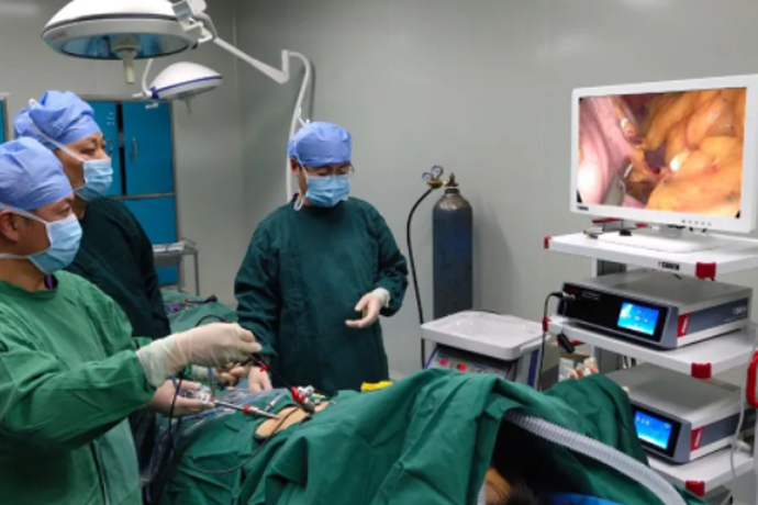
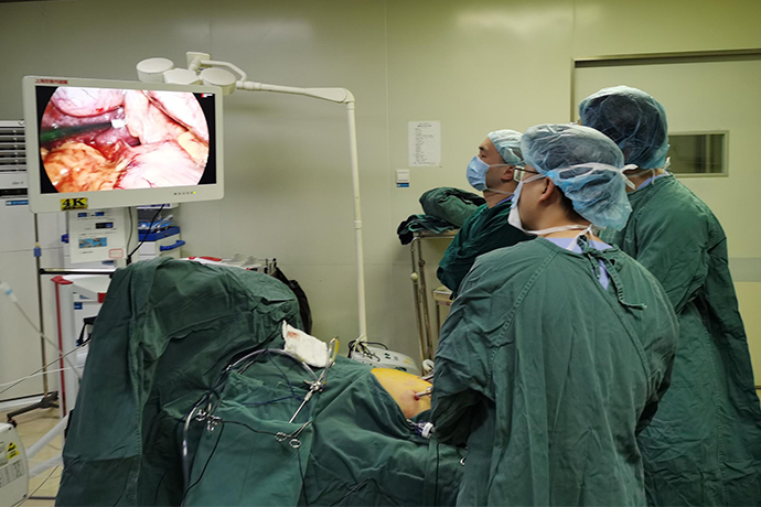
[Laparoscopic Hepatobiliary Surgery] Spleen Tumor Surgery
Release time: 11 Apr 2023 Author:Shrek
The spleen is an important lymphoid organ in the human body. It is located under the diaphragm and is protected by the surrounding bones. Therefore, the early symptoms of spleen tumors are not obvious, and it is not easy to be discovered by people, thus delaying the treatment of the disease. The symptoms of splenic tumors are related to the nature, location, size and degree of splenomegaly of the tumor. The common clinical symptoms are:

Clinical symptoms
1. Splenomegaly is mostly accompanied by symptoms of discomfort, pain and compression in the upper left abdomen, such as abdominal distension, nausea, constipation, and dyspnea.
2. There is a certain relationship between hypersplenism and splenomegaly, but the symptoms are not proportional to the degree of splenomegaly. For unexplained hypersplenism accompanied by splenomegaly, the existence of tumors, especially hemangiomas, should be highly suspected.
3. Systemic symptoms are more common in malignant tumors of the spleen, manifested as low-grade fever, anemia, fatigue, general malaise, weight loss, and cachexia.
4. Spontaneous rupture of splenic tumors is rare clinically, manifested as sudden abdominal pain, peritonitis, hemorrhagic shock and even death. Splenic rupture accompanied by early metastasis is the worst prognostic factor. Spontaneous splenic rupture is secondary to hemophagocytic syndrome, hemangiopericytoma, serous multiple hemangioma, and T-cell leukemia. Some of them may be associated with peritoneal seeding metastasis, which is more common in spontaneous rupture of splenic hemangioma and angiosarcoma.
Splenic tumors are very rare, and benign tumors are more common than malignant tumors.
Benign hemangiomas are the most common (50%), followed by lymphangiomas and hamartomas.
Malignant lymphoma is the most common malignant tumor (70%~80%), followed by metastatic tumor, angiosarcoma, and fibrous histiocytic sarcoma.
The CT features of splenic tumors are relatively unobvious, and the diagnosis should be compared.
Disease classification
The classification of splenic tumors is relatively complicated, and can be divided into the following five types according to the source of tissue components: (1) Tumor-like lesions: including non-parasitic cysts and hamartomas. (2) Vascular tumors: divided into benign and malignant. (3) Lymphoid neoplasms: Hodgkin's disease, non-Hodgkin's disease, lymphoid lesions. (4) Non-lymphoid tumors: including lipoma, malignant fibroblastoma, malignant teratoma, etc. (5) Tumor-like lesions: such as traumatic cystic pseudotumor, inflammatory pseudotumor, etc. Splenic tumors are more benign than malignant. Splenic cysts are the most common in benign tumors, and malignant lymphomas are more common in malignant tumors.
Splenic tumors are divided into the following types according to the origin: (1) Vascular origin. (2) Source of lymphoid tissue. (3) Source of embryonic tissue. (4) Source of nervous tissue. (5) Other sources of mesenchymal tissue: such as fibrous tissue, adipose tissue and smooth muscle tissue.
Surgical procedure
After general anesthesia, the patient should take a 40°right decubitus position with a head height of 30°, so that the omentum, stomach, and colon can be kept away from the spleen under the action of gravity, and a good surgical field of view can be obtained.
A 10 mm incision was made on the upper edge of the umbilical cord and a pneumoperitoneum was established. The pneumoperitoneum pressure was maintained at 14 mmHg. A 30° laparoscope was inserted to explore the abdominal cavity.
Cannula position On the midline of the left clavicle, 2 cm below the spleen, a 12 mm cannula is placed as the main operation hole (for the surgeon to use ultrasonic scalpel, Ligasure and linear cutting closure); a 5 mm cannula is placed under the xiphoid process Cannula, as an auxiliary operation hole (for the surgeon to use grasping forceps); a 5 mm cannula was placed at 2 cm below the splenic edge of the left anterior axillary line, as an auxiliary operation hole (for the first assistant to use laparoscopic grasping forceps and suction device).
Free spleen When resecting the lower pole of the spleen, it is necessary to free the splenocolic ligament, part of the splenic-gastric ligament, and the spleen-renal ligament; when resecting the upper level, it is necessary to free the splenic-diaphragmatic ligament and the corresponding splenic-gastric ligament (thick short gastric vessels can be coagulated with Ligasure) and spleen renal ligament.
Treatment of splenic hilus branch vessels: One pole of the spleen needs to be resected along the splenic hilum, and the splenic branch arteries and veins are freed at the splenic hilum. Small blood vessels can be directly coagulated with Ligasure (<5 mm), and large blood vessels can be clamped and then cut with synthetic clips. At the same time of dissociation, observe the ischemic area of the spleen. If it does not reach the resection range, continue to dissociate toward the splenic hilus (at this time, be careful not to damage the splenic artery and vein) until the ischemia range reaches the resection range.
Partial resection of the spleen Cut off the spleen 1 cm inside the ischemic area (Ultrasonic knife, combined with Ligasure), use bipolar electrocoagulation to stop bleeding from the wound, and cover the wound with hemostatic gauze. Finally, the specimen was bagged, cut into pieces and taken out, and a drainage tube was placed in the splenic fossa to complete the operation.
1. Probe the abdominal cavity, open the omentum sac until the hilum of the spleen, and pre-treat some short gastric vessels.
2. Separate the pancreaticogastric ligament; suspend the greater curvature of the gastric body.
3. Separate the adhesive omentum tissue in the splenic hilum area to expose the splenic hilum.
4. Separation of adhesions at the inferior pole of the spleen and the splenocolic ligament.
5. Separation and treatment of blood vessels at the inferior pole of the spleen.
6. Support the lower pole of the spleen, separate the lower part of the spleen-renal ligament and the space behind the hilum of the spleen.
7. Separate the splenic artery at the upper edge of the pancreas and ligate it with No. 7 silk thread.
8. Open the retroperitoneum on the inner side of the capsule of the upper pole of the spleen above the tail of the pancreas, and gradually separate the space between the spleen and stomach fundus upward along the direction of the fundus of the stomach, and deal with the remaining short gastric vessels.
9. Lift the upper pole of the spleen, separate the upper part of the spleen diaphragmatic ligament and spleen renal ligament, and join the lower part.
10. Use golden fingers to go through the tunnel behind the splenic hilum and tie the splenic pedicle with No. 7 silk thread.
11. Use golden fingers to penetrate the tunnel behind the splenic pedicle, and use Endo-GIA blue nails or white nails to close and separate the splenic pedicle.
12. Check the stump of the splenic pedicle and strengthen it with titanium clips.
13. Separate the residual splenorenal ligament.
14. The specimen is bagged and the specimen is taken out through the enlarged umbilical mirror hole.
15. Re-enter the mirror to check whether there is blood oozing from the wound, whether there is damage to the stomach wall, and whether the closed stump is reliable.
16. Place the drainage tube in the spleen fossa and spleen pedicle section.
17. Removal of the gastric suspension wire.
18. Remove the pneumoperitoneum and suture the incision.
Postoperative pathology
General: 1 spleen specimen, about 15.0 cm × 8.2 cm × 3.0 cm in size, part of the spleen tissue was severed, the cross-section was bean paste red, no hematoma was seen in the parenchyma, and no obvious gray-white mass was seen in multiple sections.
Pathological diagnosis: chronic congestive splenomegaly.
Summarize
Surgery is safe and feasible for experienced minimally invasive surgeons. At the same time, partial laparoscopic splenectomy has the following advantages: less intraoperative blood loss and shorter operation time; fast postoperative recovery and fewer complications; the important role of the spleen in immunity and anti-tumor is preserved; the cosmetic effect is good and patients are easy to accept . In view of this, laparoscopic partial splenectomy may partially replace total splenectomy and become a new method of minimally invasive surgery for spleen diseases.

- Recommended news
- 【General Surgery Laparoscopy】Cholecystectomy
- Surgery Steps of Hysteroscopy for Intrauterine Adhesion
- [Otolaryngology Otoscopic Section] Excision of Cholesteatoma in the External Auditory Canal
- [Otolaryngology Nasal Endoscopy] How to Treat Recurrent Rhinitis
- [ENT Surgery: Nasal Endoscopy] Endoscopic Treatment of Nasal Polyps