【Gynecology Laparoscopy】Have You Mastered These Basic Essentials?
Release time: 08 Jun 2021 Author:Shrek
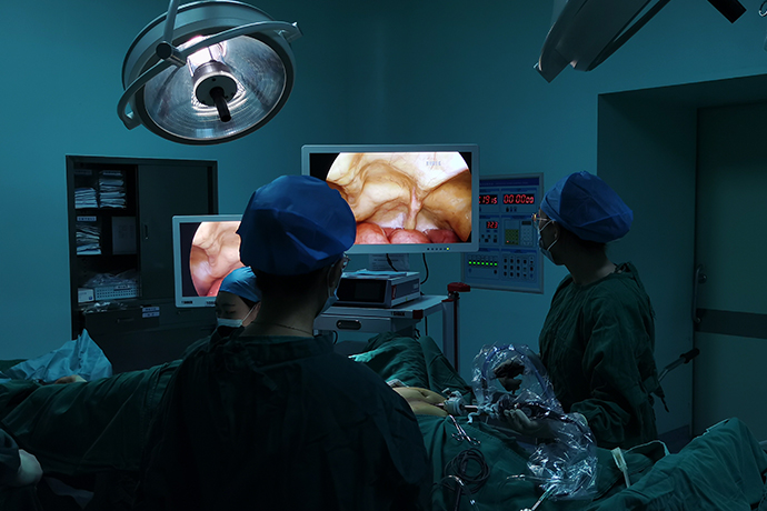
Laparoscopic surgery is one of the better widely used surgical methods in gynecology nowadays. It has the obvious characteristics of minimally invasive, small scars, and quick recovery. It has become the three basic techniques of gynecological surgery together with open surgery and vaginal surgery.
Formation of artificial pneumoperitoneum
For most laparoscopic surgeries, using pneumoperitoneum to fully expose the surgical field is the first step to successful surgery.
(1) Selection of puncture site
The umbilicus is a natural scar, and this part (above and below the umbilicus) is the best place for puncture.
(2) Puncture method
Lift the skin on both sides, cut the umbilical skin at the puncture site, used a pen holding pneumoperitoneum needle, and make the puncture at 90° with the wrist against the abdominal wall.
Tips:
There are two times empty feeling during the puncture process, the first time is to penetrate the anterior rectus sheath, and the second time is to penetrate the peritoneum into the abdominal cavity.
After the needle is inserted into the abdomen, the tail of the needle is connected to a small syringe containing saline. After the second time of empty feeling, the saline will automatically enter the abdominal cavity slowly due to the negative pressure in the abdominal cavity, and the liquid level will drop.
Hemostasis method
(1) Mechanical hemostasis
Clamping hemostasis: It is used to block medium-sized blood vessels. The two ends of the blood vessel can be clamped, and then the blood vessel can be quarantine from the middle. Including titanium clips and ligature locks (Hem-o-lok), etc.
Suture to stop bleeding: through suture
Compression to stop bleeding: gauze or equipment
(2) Electric energy to stop bleeding
High-frequency electrosurgical hemostasis: single-stage electrosurgical or bipolar electrocoagulation system.
Ultrasonic hemostatic knife: It can coagulate and close relatively large lumens and blood vessels without producing obvious smoke. The surgical field of vision is clear, and fine operations can be performed in small spaces and deep in the surgical field.
Ligasure Vascular Closure System: It can close relatively thick blood vessels with fast closing speed, no smoke, and does not affect the surgical field of vision.
(3) Drugs to stop bleeding
Thrombin: Thrombin is more commonly used in hemostatic drugs. Thrombin is a highly specific serine proteolytic enzyme that plays an important role in regulating platelet aggregation and blood clotting in hemostasis.
Gel sponge etc.
Indications and contraindications
(1) The best indication
Laparoscopic surgery is usually the preferred surgical method, which can effectively confirm the diagnosis and carry out the corresponding treatment.
1. Acute abdomen: such as ectopic pregnancy, torsion of ovarian cyst pedicle, rupture of ovarian cyst, etc.
2. Accessory masses: such as benign ovarian tumors, mesangial cysts, and accessory inflammatory masses.
3. Endometriosis.
4. Chronic pelvic pain.
5. Infertility.
6. Others: such as foreign bodies in the pelvic and abdominal cavity, uterine perforation, etc.
(2) Selective indications
Laparoscopy is an alternative surgical method.
1. Uterine fibroids: perform uterine fibroids or hysterectomy under laparoscopy.
2. Adenomyosis: Perform adenomyosis lesion resection or hysterectomy under laparoscopy.
3. Early endometrial cancer, early cervical cancer, early borderline ovarian tumor and epithelial ovarian cancer (ovarian cancer), etc.: Perform tumor staging and re-staging surgery under laparoscopic surgery, as well as fertility-preserving surgery for early cervical cancer.
4. Pelvic floor dysfunction disease: Perform laparoscopic pelvic floor reconstruction surgery.
5. Abnormal development of reproductive organs; artificial vaginoplasty, etc.
6. Accessory mass during pregnancy.
7. Other diseases that require removal of the uterus and/or accessories.
(3) Absolute contraindications
1. Severe heart and cerebrovascular diseases and pulmonary insufficiency.
2. Severe coagulation dysfunction and blood disease.
3. Diaphragmatic hernia.
(4) Relative contraindications
1. Extensive adhesions in the pelvic and abdominal cavity.
2. Huge attachment masses.
3. Intramuscular uterine fibroids are large in size (diameter ≥10cm) or more in number (≥4) and require preservation of the uterus.
4. Late or widely metastatic gynecological malignant tumors.
Treatment during operation
(1) Preoperative preparation
1. Preoperative examination: hematuria routine, blood type (including Rh blood type), clotting time, liver and kidney function, five items of hepatitis B, hepatitis C antibodies, syphilis and HIV, electrocardiogram, chest X-ray, B-ultrasound. Cardiopulmonary function, echocardiography, cervical cytology, gynecological tumor markers, vaginal secretions and pelvic and abdominal cavity MRI and CT examinations should be completed when necessary.
2. Skin preparation: In accordance with the procedures of abdominal and perineal surgery, pay special attention to the cleanliness of the umbilicus.
3. Vaginal preparation: Vaginal douche before surgery as appropriate.
4. Intestinal preparation: ld oral laxative before surgery, enema or cleaning enema if necessary, fasting for more than 6h before surgery.
5. Bladder preparation: emptying the bladder, catheterization or indwelling catheter.
(2) Postoperative treatment
1. After the operation, determine the time of eating, getting out of bed, and indwelling catheter.
2. Carefully monitor changes in symptoms and signs such as body temperature, surgical incision, subcutaneous hematoma or emphysema, exhaust or defecation, and timely detect and deal with postoperative complications. If necessary, monitor peripheral blood and other indicators.
3. Postoperative upper abdomen (especially diaphragmatic ribs) and shoulder pain generally do not require special treatment, and oral analgesics can be given if necessary.
Basic requirements
1. Exposure of the surgical field: the intestine can be moved to the upper abdomen with the head low, which can fully expose the pelvic organs. With the help of the uterine lifter to swing the uterus, the tissue structure of the front and back of the uterus can be exposed. The surgeon or assistant uses surgical instruments to move in the abdominal cavity. Or lift the corresponding tissues and organs, and separate adhesions if necessary to achieve the purpose of effectively exposing the surgical field.
2. Tissue cutting: Use electrosurgical knife, scissors or ultrasonic knife to separate and cut the tissue.
3. Tissue separation: According to the intraoperative conditions, sharp (using separation forceps, scissors, electric knife, etc.), blunt (using a stripping stick, aspirator or "peanut" stripper) or hydraulic separation can be selected.
4. Knotting: Knotting outside the body and inside the abdominal cavity.
5. Hemostasis: The commonly used methods of hemostasis include electrocoagulation, stitching and clamping of blood vessels, and gauze compression.
6. Organ and tissue repair: suture methods are often used.
7. Tissue or specimen removal: It can be taken out directly through the puncture cannula: the uterus or uterine fibroids can be cut out piece by piece with a tissue pulverizer, or taken out through the vagina. It is recommended that tissue specimens be taken out using specimen bags.
Intraoperative monitoring
Intraoperative comprehensive monitoring of blood pressure, respiration, heart rate and other vital signs: monitoring of pulse, blood oxygen saturation and CO2 partial pressure: monitoring of intraoperative blood loss at the same time.
Surgical grade
Level 1 surgery
1. Laparoscopy
2. Fallopian tube sterilization
3. Pelvic and abdominal tissue biopsy
4. Medicine injection for tubal pregnancy
5. Mild pelvic adhesion lysis
6. Early peritoneal endometriosis lesions cauterization
Level 2 surgery
1. Fenestration of tubal pregnancy
2. Salpingectomy
3. The function of fallopian tube is eliminated
4. Removal of mesangial and ovarian crown cysts
5. Simple ovarian cystectomy
6. Partial or wedge resection of ovary
7. Ovarian perforation
8. Ovarian (or) adnexectomy (except for severe adhesions)
9. Removal of free foreign bodies in abdominal cavity
10. Uterine round ligament suspension
Level 3 surgery
1. Total hysterectomy and accessory resection or laparoscopic assisted vaginal hysterectomy (LAVH)
2. Subtotal hysterectomy
3. Uterine myomectomy
4. Removal of ovarian endometriotic cysts or excision of accessories 5. Resection of adenomyosis lesions
5. Resection of scar pregnancy lesions after cesarean section
6. Surgical treatment of pelvic encapsulated effusion
7. Release of moderate and severe pelvic adhesions
8. Incision and drainage of pelvic abscess
9. Uterine repair
10. Remnant horn hysterectomy
11. Cut off the uterosacral nerve
12. High uterosacral ligament suspension
13. Resection of attachments with severe adhesions
Level 4 surgery
1. Total hysterectomy with uterine volume ≥12 gestational weeks
2. Resection of deep infiltrating endometriosis lesions
3. Total hysterectomy with severe endometriosis
4. Extensive hysterectomy
5. Pelvic lymph node resection
6. Abdominal para-aortic lymph node resection
7. Omentumectomy
8. Extensive cervical resection
9. Presacral neurotomy
10. Tubal anastomosis
11. Uterine and (or> vaginal sacral fixation
12. Bladder neck suspension
13. Vaginoplasty
14. Bicornuate Hysteroplasty
15. Laparoscopic surgery in the second trimester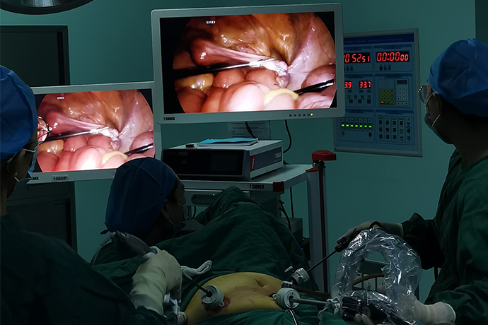
- Recommended news
- 【General Surgery Laparoscopy】Cholecystectomy
- Surgery Steps of Hysteroscopy for Intrauterine Adhesion
- [Percutaneous Endoscopic Lumbar Discectomy] Percutaneous endoscopic lumbar discectomy for lumbar disc herniation
- [Urology Cystoscopy Section] Cystoscopic Laser Lithotripsy
- [Laparoscopic Urology] Laparoscopic Radical Right Nephrectomy