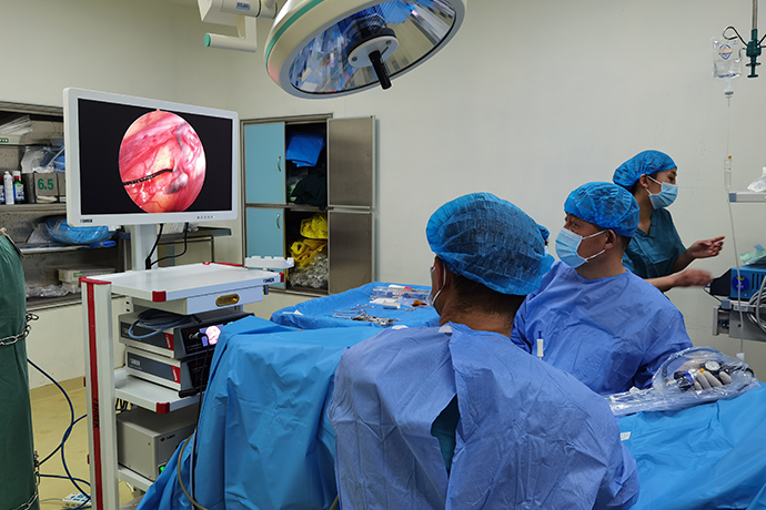
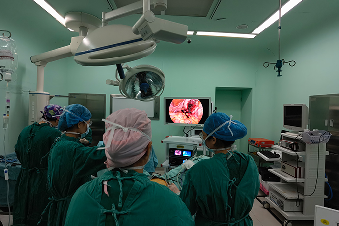
【Gynecological Laparoscopy】Hysterectomy
Release time: 17 Jun 2021 Author:Shrek
"The clinical application of laparoscopic surgery is not just a change in surgical methods, but an event of great significance to promote the change of concepts of surgeons." Professor Liu Yan mentioned in the report: Application of laparoscopic surgery Gynecologists have a great discussion about the practice of various minimally invasive procedures and the concept of "minimally invasive". The most clear manifestation is that the application of laparoscopic surgery has prompted doctors accustomed to transabdominal surgery to change the traditional concepts of "large incision" and "large exposure", and try to be "minimally invasive" on surgical incisions, and then perform multiple operations with laparoscopic surgery. The surgical method of the hole is comparable to the competition. On the other hand, laparoscopic surgery has also prompted people to re-emphasize transvaginal surgery without abdominal wall incisions in gynecological surgery.
Laparoscopic total laproseopie hysterectomy (TLH), TLH is a kind of laparoscopic cutting of ligaments, blood vessels and vaginal wall tissues around the uterus to completely free the uterus and then remove it from the vagina and suture it under laparoscopic surgery Surgical method of vaginal stump.

Indications
1. Benign lesions of the uterus: such as uterine fibroids, adenomyosis, dysfunctional uterine bleeding, etc. who need to remove the uterus;
2. Early uterine malignant tumors, such as cervical carcinoma in situ, cervical epithelial or endometrial dysplasia and other patients who are suitable for total hysterectomy.
3. The uterus should not exceed the size of 16 weeks of pregnancy.
Severe pelvic adhesions should be considered as a contraindication for laparoscopic surgery.
Surgical steps and technical points
1. Deal with the round ligament of the uterus and the proper ligament of the ovary
For those who need to preserve the ovaries, the uterine device is pushed to one side of the uterus, and at the same time, the round ligament is expanded at 2cm near the uterine horn, and the round ligament is cut off by electrocoagulation at 2cm or the middle part of the uterine horn. Then, the fallopian tube was electrocoagulated at a distance of about 2cm from the uterine horn, and the fallopian tube was cut off. Then the proper ligament of the ovary was electrocoagulated at 1 cm from the uterine horn. Separate the middle section of the broad ligament, apply bipolar electrocoagulation forceps glue, and then cut the ligament and tissue with an ultrasonic knife. If the ligament is thickened and hardened, especially in endometriosis, the thickened tissue should be fully electrocoagulated. If the coagulation is insufficient, bleeding may occur and affect the operation, and the cutting should be close to the ovarian side.
Step 1. Cut off the round ligament
Step 2. Ovarian vessel cut
2. Treatment of uterine blood vessels After treatment of the round ligament, broad ligament and proper ovarian ligament, open 2cm next to the cervix junction, open the posterior lobe of the broad ligament about 1.5cm above the uterosacral ligament, separate the connective tissue, and expose the uterine artery. The uterine artery is blocked by bipolar electrocoagulation and dehydration. Avoid damaging the ureter: (1) Cut the uterine blood vessels on the anterior and posterior sides; (2) Choose the ascending branch of the uterine artery for electrocoagulation; (3) Try to shorten the electrocoagulation time. Short-term and repeated electrocoagulation are better than long-term, Continuous coagulation. At the same time, the assistant pushes the uterus from the vagina to the head at the critical moment to keep the uterine blood vessels away from the ureter; (4) During the free exposure of the uterine artery, the uterine artery should be fully freed as much as possible. If the adjacent ureter can be exposed and pushed at the same time Leaving the uterine artery is the most accurate measure to avoid ureteral damage.
Step 3. Cut off the posterior lobe of the broad ligament
3. Open the bladder and peritoneum reflex, push the bladder down
Cut the broad ligament from the broken end of the round ligament toward the cervix to about 5mm below the bladder-uterine peritoneum junction. Use grasping forceps to clamp the bladder and fold the peritoneum and anterior wall. At the same time, use the uterine lift to push the uterus, cervix and vagina toward the head. At the upper part of the junction, push the bladder down along the edge of the uterine cup, and push it down to about 2cm below the outer cervix. In case of bleeding, bipolar electrocoagulation can be used to stop the bleeding. Slow cutting when using an ultrasonic knife can achieve a good hemostatic effect.
Step 4. Cut off the bladder, uterus and peritoneum
Step 5. Dissecting the bladder-uterine space
Step 6. Uterine artery cut
Step 7. Circular vaginectomy
Step 8. Closing the vaginal cuffs
4. Cut the fornix and remove the uterus
Expose the fornix, cut the fornix at the upper edge of the cup, and then cut the lateral fornix and posterior fornix in turn, and the uterus is removed from the body. Subtotal hysterectomy patients do not need to cut the fornix or cervix.
5. Turn off the blockage and cervical stump
The use of No. 0 and No. 2-0 needles can absorb tension suture. The suture method can be intermittent suture, intermittent "8" suture, continuous buckle suture and so on. The continuous penetration suture has a good effect on the bleeding of the vagina.
6. Check the pelvic and abdominal cavity
After closing the fornix, use a laparoscope to examine the pelvic cavity, flush and suck the blood clots and debris, flushing helps to find some small bleeding, apply bipolar coagulation to further stop the bleeding, and suture if necessary to stop the bleeding. Check the activity of ureter, normal peristalsis and no dilatation can exclude ureteral injury, only peristalsis is not confirmed. For cases with a lot of lavage fluid and large surgical wounds, in principle, it is recommended to indwell a pelvic drainage tube after the operation.
Precautions after operation:
1. Indwelling the urinary catheter for 24 to 48 hours after the operation; observe the urine color and urine volume.
2. Use antibiotics for 2 to 3 days after surgery, and get out of bed for 12 to 24 hours.
Surgical skills and prevention of complications
1. To raise the uterus cup, be sure to lift the anterior and posterior fornix of the vagina, and lift the uterus;
2. When the isthmus of the fallopian tube, the proper ligament of the ovary and the round ligament are cut, do not get too close to the corner of the uterus, otherwise there will be more bleeding and difficulty in hemostasis;
3. Cut open the bladder and peritoneum at the loose part and fold it, not too low, so as not to damage the bladder; push the bladder down to 2cm below the outer cervix, there is room for suture the vaginal stump, it is not easy to sew to the bladder.
4. When pushing down the bladder, pay attention to electrocoagulation and incision of the bladder and cervix ligament, instead of blindly pushing down to cause bleeding;
5. When using bipolar coagulation or Ligasure to coagulate the uterine blood vessels, it must be close to the cervix to avoid ureteral damage. If necessary, dissect the free ureter.
6. When you cut the vaginal fornix with a ring, you can use a monopolar or ultrasonic knife. When using a unipolar electric hook, you should pay attention to controlling the coagulation time to avoid excessive postoperative leakage. When incising the vagina on both sides of the vagina, pay attention to the vaginal blood vessels. Electrocoagulation first
7. Do not suture the vaginal stump too tightly, otherwise it will easily become necrotic and fall off, causing excessive vaginal exudation after surgery, and even formation of polyps;
8. After the suture, check the wound surface. If bleeding occurs, use bipolar coagulation to stop the bleeding. After the operation, use a speculum to check whether the vaginal stump is seamlessly closed, and whether there are lacerations on the vaginal wall and vaginal opening.

Laparoscopic total laproseopie hysterectomy (TLH), TLH is a kind of laparoscopic cutting of ligaments, blood vessels and vaginal wall tissues around the uterus to completely free the uterus and then remove it from the vagina and suture it under laparoscopic surgery Surgical method of vaginal stump.

Indications
1. Benign lesions of the uterus: such as uterine fibroids, adenomyosis, dysfunctional uterine bleeding, etc. who need to remove the uterus;
2. Early uterine malignant tumors, such as cervical carcinoma in situ, cervical epithelial or endometrial dysplasia and other patients who are suitable for total hysterectomy.
3. The uterus should not exceed the size of 16 weeks of pregnancy.
Severe pelvic adhesions should be considered as a contraindication for laparoscopic surgery.
Surgical steps and technical points
1. Deal with the round ligament of the uterus and the proper ligament of the ovary
For those who need to preserve the ovaries, the uterine device is pushed to one side of the uterus, and at the same time, the round ligament is expanded at 2cm near the uterine horn, and the round ligament is cut off by electrocoagulation at 2cm or the middle part of the uterine horn. Then, the fallopian tube was electrocoagulated at a distance of about 2cm from the uterine horn, and the fallopian tube was cut off. Then the proper ligament of the ovary was electrocoagulated at 1 cm from the uterine horn. Separate the middle section of the broad ligament, apply bipolar electrocoagulation forceps glue, and then cut the ligament and tissue with an ultrasonic knife. If the ligament is thickened and hardened, especially in endometriosis, the thickened tissue should be fully electrocoagulated. If the coagulation is insufficient, bleeding may occur and affect the operation, and the cutting should be close to the ovarian side.
Step 1. Cut off the round ligament
Step 2. Ovarian vessel cut
2. Treatment of uterine blood vessels After treatment of the round ligament, broad ligament and proper ovarian ligament, open 2cm next to the cervix junction, open the posterior lobe of the broad ligament about 1.5cm above the uterosacral ligament, separate the connective tissue, and expose the uterine artery. The uterine artery is blocked by bipolar electrocoagulation and dehydration. Avoid damaging the ureter: (1) Cut the uterine blood vessels on the anterior and posterior sides; (2) Choose the ascending branch of the uterine artery for electrocoagulation; (3) Try to shorten the electrocoagulation time. Short-term and repeated electrocoagulation are better than long-term, Continuous coagulation. At the same time, the assistant pushes the uterus from the vagina to the head at the critical moment to keep the uterine blood vessels away from the ureter; (4) During the free exposure of the uterine artery, the uterine artery should be fully freed as much as possible. If the adjacent ureter can be exposed and pushed at the same time Leaving the uterine artery is the most accurate measure to avoid ureteral damage.
Step 3. Cut off the posterior lobe of the broad ligament
3. Open the bladder and peritoneum reflex, push the bladder down
Cut the broad ligament from the broken end of the round ligament toward the cervix to about 5mm below the bladder-uterine peritoneum junction. Use grasping forceps to clamp the bladder and fold the peritoneum and anterior wall. At the same time, use the uterine lift to push the uterus, cervix and vagina toward the head. At the upper part of the junction, push the bladder down along the edge of the uterine cup, and push it down to about 2cm below the outer cervix. In case of bleeding, bipolar electrocoagulation can be used to stop the bleeding. Slow cutting when using an ultrasonic knife can achieve a good hemostatic effect.
Step 4. Cut off the bladder, uterus and peritoneum
Step 5. Dissecting the bladder-uterine space
Step 6. Uterine artery cut
Step 7. Circular vaginectomy
Step 8. Closing the vaginal cuffs
4. Cut the fornix and remove the uterus
Expose the fornix, cut the fornix at the upper edge of the cup, and then cut the lateral fornix and posterior fornix in turn, and the uterus is removed from the body. Subtotal hysterectomy patients do not need to cut the fornix or cervix.
5. Turn off the blockage and cervical stump
The use of No. 0 and No. 2-0 needles can absorb tension suture. The suture method can be intermittent suture, intermittent "8" suture, continuous buckle suture and so on. The continuous penetration suture has a good effect on the bleeding of the vagina.
6. Check the pelvic and abdominal cavity
After closing the fornix, use a laparoscope to examine the pelvic cavity, flush and suck the blood clots and debris, flushing helps to find some small bleeding, apply bipolar coagulation to further stop the bleeding, and suture if necessary to stop the bleeding. Check the activity of ureter, normal peristalsis and no dilatation can exclude ureteral injury, only peristalsis is not confirmed. For cases with a lot of lavage fluid and large surgical wounds, in principle, it is recommended to indwell a pelvic drainage tube after the operation.
Precautions after operation:
1. Indwelling the urinary catheter for 24 to 48 hours after the operation; observe the urine color and urine volume.
2. Use antibiotics for 2 to 3 days after surgery, and get out of bed for 12 to 24 hours.
Surgical skills and prevention of complications
1. To raise the uterus cup, be sure to lift the anterior and posterior fornix of the vagina, and lift the uterus;
2. When the isthmus of the fallopian tube, the proper ligament of the ovary and the round ligament are cut, do not get too close to the corner of the uterus, otherwise there will be more bleeding and difficulty in hemostasis;
3. Cut open the bladder and peritoneum at the loose part and fold it, not too low, so as not to damage the bladder; push the bladder down to 2cm below the outer cervix, there is room for suture the vaginal stump, it is not easy to sew to the bladder.
4. When pushing down the bladder, pay attention to electrocoagulation and incision of the bladder and cervix ligament, instead of blindly pushing down to cause bleeding;
5. When using bipolar coagulation or Ligasure to coagulate the uterine blood vessels, it must be close to the cervix to avoid ureteral damage. If necessary, dissect the free ureter.
6. When you cut the vaginal fornix with a ring, you can use a monopolar or ultrasonic knife. When using a unipolar electric hook, you should pay attention to controlling the coagulation time to avoid excessive postoperative leakage. When incising the vagina on both sides of the vagina, pay attention to the vaginal blood vessels. Electrocoagulation first
7. Do not suture the vaginal stump too tightly, otherwise it will easily become necrotic and fall off, causing excessive vaginal exudation after surgery, and even formation of polyps;
8. After the suture, check the wound surface. If bleeding occurs, use bipolar coagulation to stop the bleeding. After the operation, use a speculum to check whether the vaginal stump is seamlessly closed, and whether there are lacerations on the vaginal wall and vaginal opening.

- Recommended news
- 【General Surgery Laparoscopy】Cholecystectomy
- Surgery Steps of Hysteroscopy for Intrauterine Adhesion
- [ENT Surgery: Nasal Endoscopy] Endoscopic Treatment of Nasal Polyps
- [Otolaryngology Nasal Endoscopy] Methods and Precautions for Nasal Bleeding Control under Nasal Endoscopy
- [Orthopedic UBE Section] Four Years of Evolution of UBE Technology: From Lumbar Fusion to the "Forbidden Zone" of Thoracic and Cervical Spine, and the Unignorable "Hydraulic Pressure Crisis"