Breast Duct Endoscope
Release time: 11 Aug 2021 Author:Shrek
Minimally Invasive Endoscope
Maybe you have heard of gastroscopy, colonoscopy, laparoscopy, have you heard of breast ductscopy? Let’s talk about the breast duct endoscope with the smallest and smallest diameter in the endoscope today!
An artifact to find out the cause of nipple discharge
Nipple discharge is one of the common symptoms in breast clinics, and it is also easier for patients to pay attention to. It is one of the main reasons for about 10% of clinical patients to come to see a doctor. Nipple discharge is mainly caused by physiological factors, systemic diseases, and breast diseases. 5%-10% of breast cancer patients have nipple discharge. Clinically, about 1% of breast cancers have nipple discharge as the first symptom or the only symptom. Because the change of malignant disease can easily cause bloody discharge, clinical patients with bloody discharge should be more alert to the possibility of malignant disease. If a male patient has nipple discharge, it should not be taken lightly.
What is a breast duct endoscope?
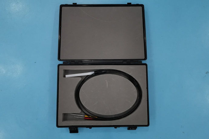
Breast duct endoscope is called ultra-fine fiber optic mammary duct endoscope, its lens diameter is only 0.7mm, 0.3mm working injection channel, tortuosity rate of 40°, it can perform any branch at all levels, including intraoperative surgery, and inject liquid medicine at the same time. treatment. Designed according to the characteristics of the breast of the Asian race. The endoscope body is made by pouring waves as a whole, which is safe and durable. Field of view: ≥70%, (not counted on the line), the viewing angle is 0°, it can penetrate into the six-level breast duct without error, and the number of uses is more than 200 times. Pixels: 6000 pixels, with reinforced steel wire, not easy to break. The observation range is from the opening of the nipple duct to the distal end of 5-6cm, and the maximum insertion depth is (4.5±1)cm on average, which can basically meet the clinical needs. It can adapt to various disinfection measures and is very durable, reliable and safe. Working distance is 6cm. Working hole: 0.3mm (sus tube) can be equipped with biopsy, cell rotary biopsy instrument, with hook positioning needle before operation, dredging guide wire.
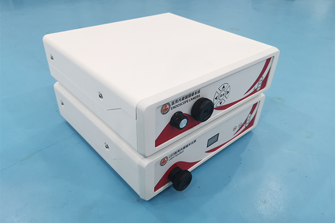
Through the breast duct endoscope, the doctor can see the lesions in the duct at a glance. It is a cost-effective clinical diagnosis technique, and it has become the first choice for diagnosis of the cause of nipple discharge. Since 2009, the clinical application of breast duct endoscopy has replaced the past breast duct angiography.
Why do we need a breast duct endoscope?
1. Part of the nipple discharge is only caused by chronic inflammation in the breast duct. After a clear diagnosis, only follow-up or regular drug washing under the ductoscope is required to avoid unnecessary surgery.
2. For intraductal space-occupying lesions, breast ductscopy can repeatedly observe them and make precise positioning. Provide surgical basis, ensure the accuracy of the surgical location, and reduce the scope of surgery.
3. For early breast cancer whose lesions are still confined to the duct, breast ductscopy is the earliest examination method to find its existence. Once discovered, early diagnosis and early treatment can be achieved.
Who needs breast endoscopy?
Common breast diseases, such as intraductal papilloma, duct dilatation, mastitis, breast hyperplasia, and even breast cancer can all be manifested as nipple discharge.
Generally speaking, patients with bloody or yellow discharge are more likely to have neoplastic lesions in the ducts. Patients with pale yellow clear or turbid discharge have a greater possibility of chronic inflammation in the breast ducts. In patients with clear watery discharge, there is a greater possibility of milder breast duct inflammation such as duct dilatation.
If there is nipple discharge, you need to see a doctor in time. After consultation and physical examination by a breast specialist, you will decide whether you need a breast ductscopy based on the specific situation.
How to check by breast endoscope?
1. The patient is lying flat all the way, in a more comfortable position. The patient is fully awake and can see the inspection process on the display with the doctor. The inspection process takes about 3-5 minutes. After the inspection, you can leave and you can get the report on the spot.
2. At the beginning of the examination, after disinfection, the doctor will use a probe to dilate the breast duct, then change the ductscope to enter, and inject anesthetics, physiological saline and other drugs through the working channel to flush. Through the skilled operation of the doctor, the whole line can be clearly seen lesions in the breast ducts.
3. If new organisms in the catheter, stenosis of the tube wall, bleeding, or suspected lesions are found, the doctor will adjust the lens angle and the brightness of the light source, repeatedly observe carefully, record the specific location, and take pictures on the spot to provide for the next surgical treatment diagnosis basis and precise positioning.
Does breast endoscopy hurt?
The breast duct is indeed very thin, but the breast duct lens is also very thin. Moreover, the breast ducts of patients with discharge usually have different degrees of dilation, so there is no pain or only slight pain.
What should I pay attention to before and after breast endoscopy?
1. First relax the inspection mentality. Breast endoscopy is a non-invasive examination. It enters the existing physiological channel and will not cause damage to the human body;
2. The nipple is obviously red and swollen, and breast ductscopy should be avoided during acute inflammation;
3. Try to avoid breast ductscopy during menstruation;
4. Patients with serious medical diseases need to undergo breast endoscopy after their condition is stable;
5. Patients with fever caused by any reason on the day of the examination cannot undergo breast endoscopy;
6. No bathing for 24 hours after the inspection.
Introduction to Professional Edition
Breast endoscopy can directly observe the changes of the breast duct epithelium, and can perform qualitative and localized diagnosis of the small lesions in the breast ducts with bloody or serous discharge of the nipple and no mass in the breast. It is an important clinical examination method for breast diseases.
The scope of application of breast ductscopy
1. All kinds of nipple discharge
Especially in patients with unilateral, single-port nipple discharge, nipple bloody discharge, and yellow discharge, the incidence of neoplastic lesions in the breast duct is about 1/3 to 1/2. In addition, patients with white or colorless discharge are also Many cases are caused by tumors in the breast ducts, which require surgical treatment after ductoscopy.
2. Masses in the areola area with nipple discharge
Most of the tumors in this area are caused by tumors in the breast ducts, which are closely related to the breast ducts. Through ductoscopy, the diseased breast ducts can be identified, so as to guide the operation, accurately remove the tumors and the diseased ducts, and reduce the chance of local recurrence after surgery. .
80% of breast cancer patients are first diagnosed with breast masses. Patients often find breast lumps inadvertently, which are mostly single, hard, with irregular edges and unsmooth surfaces. Most breast cancers are painless masses, and only a few have varying degrees of dull pain or tingling.
3. Patients with plasma cell mastitis around the areola
Plasma cell mastitis is caused by the blockage of the proximal breast duct, the accumulation of secretions, exfoliated cells, and inflammatory cells in the breast duct, which causes acute and chronic inflammation of the breast. The breast duct endoscope can lavage and collect the exfoliated cells in the breast duct, perform cytological examination, and confirm the diagnosis. At the same time, it can also flush and dredge the diseased breast ducts to achieve the purpose of drainage. In addition, if the inflammation is relatively limited, the diseased breast ducts can also be identified under a ductoscope and the diseased breast ducts can be surgically removed.
4. Mammary pain
Mammary pain is a type of breast hyperplasia, partly due to blockage of the proximal breast ducts, which makes the distal breast ducts poorly excreted and twisted. The breast duct lavage and the dredging of the breast ducts under the ductoscope can help. Make a clear diagnosis and achieve a certain therapeutic effect.
5. Skin changes
The skin changes caused by breast cancer can have a variety of signs. The most common is that the tumor invades the Cooper ligament that connects the breast skin and the deep pectoral muscle fascia. That is, there is a small depression in the breast skin, like a small dimple. If cancer cells block the lymphatic vessels, there will be "orange peel-like changes", that is, many small dot-like depressions appear on the breast skin, just like orange peels. In advanced breast cancer, cancer cells infiltrate into the skin and grow along the lymphatic vessels, gland ducts or fibrous tissue, forming scattered hard nodules in the skin around the main cancer focus, the so-called "skin satellite nodules."
6. Swollen axillary lymph nodes
More than one third of breast cancer patients admitted to major hospitals have axillary lymph node metastasis. In the early stage, there may be swollen lymph nodes in the ipsilateral axillary, and the enlarged lymph nodes are hard, scattered, and pushable. As the disease progresses, the lymph nodes gradually fuse, and adhere to and fixate with the skin and surrounding tissues. In the late stage, the metastatic lymph nodes can be felt on the clavicle and the contralateral armpit.
Breast endoscopy is an examination and treatment using human physiological channels. It is truly non-invasive and no scars. During the treatment, you can observe the images and understand the condition under the doctor's explanation. Generally, there is no need to change the dressing after the treatment.
Maybe you have heard of gastroscopy, colonoscopy, laparoscopy, have you heard of breast ductscopy? Let’s talk about the breast duct endoscope with the smallest and smallest diameter in the endoscope today!
An artifact to find out the cause of nipple discharge
Nipple discharge is one of the common symptoms in breast clinics, and it is also easier for patients to pay attention to. It is one of the main reasons for about 10% of clinical patients to come to see a doctor. Nipple discharge is mainly caused by physiological factors, systemic diseases, and breast diseases. 5%-10% of breast cancer patients have nipple discharge. Clinically, about 1% of breast cancers have nipple discharge as the first symptom or the only symptom. Because the change of malignant disease can easily cause bloody discharge, clinical patients with bloody discharge should be more alert to the possibility of malignant disease. If a male patient has nipple discharge, it should not be taken lightly.
What is a breast duct endoscope?
Breast duct endoscope is called ultra-fine fiber optic mammary duct endoscope, its lens diameter is only 0.7mm, 0.3mm working injection channel, tortuosity rate of 40°, it can perform any branch at all levels, including intraoperative surgery, and inject liquid medicine at the same time. treatment. Designed according to the characteristics of the breast of the Asian race. The endoscope body is made by pouring waves as a whole, which is safe and durable. Field of view: ≥70%, (not counted on the line), the viewing angle is 0°, it can penetrate into the six-level breast duct without error, and the number of uses is more than 200 times. Pixels: 6000 pixels, with reinforced steel wire, not easy to break. The observation range is from the opening of the nipple duct to the distal end of 5-6cm, and the maximum insertion depth is (4.5±1)cm on average, which can basically meet the clinical needs. It can adapt to various disinfection measures and is very durable, reliable and safe. Working distance is 6cm. Working hole: 0.3mm (sus tube) can be equipped with biopsy, cell rotary biopsy instrument, with hook positioning needle before operation, dredging guide wire.
Through the breast duct endoscope, the doctor can see the lesions in the duct at a glance. It is a cost-effective clinical diagnosis technique, and it has become the first choice for diagnosis of the cause of nipple discharge. Since 2009, the clinical application of breast duct endoscopy has replaced the past breast duct angiography.
Why do we need a breast duct endoscope?
1. Part of the nipple discharge is only caused by chronic inflammation in the breast duct. After a clear diagnosis, only follow-up or regular drug washing under the ductoscope is required to avoid unnecessary surgery.
2. For intraductal space-occupying lesions, breast ductscopy can repeatedly observe them and make precise positioning. Provide surgical basis, ensure the accuracy of the surgical location, and reduce the scope of surgery.
3. For early breast cancer whose lesions are still confined to the duct, breast ductscopy is the earliest examination method to find its existence. Once discovered, early diagnosis and early treatment can be achieved.
Who needs breast endoscopy?
Common breast diseases, such as intraductal papilloma, duct dilatation, mastitis, breast hyperplasia, and even breast cancer can all be manifested as nipple discharge.
Generally speaking, patients with bloody or yellow discharge are more likely to have neoplastic lesions in the ducts. Patients with pale yellow clear or turbid discharge have a greater possibility of chronic inflammation in the breast ducts. In patients with clear watery discharge, there is a greater possibility of milder breast duct inflammation such as duct dilatation.
If there is nipple discharge, you need to see a doctor in time. After consultation and physical examination by a breast specialist, you will decide whether you need a breast ductscopy based on the specific situation.
How to check by breast endoscope?
1. The patient is lying flat all the way, in a more comfortable position. The patient is fully awake and can see the inspection process on the display with the doctor. The inspection process takes about 3-5 minutes. After the inspection, you can leave and you can get the report on the spot.
2. At the beginning of the examination, after disinfection, the doctor will use a probe to dilate the breast duct, then change the ductscope to enter, and inject anesthetics, physiological saline and other drugs through the working channel to flush. Through the skilled operation of the doctor, the whole line can be clearly seen lesions in the breast ducts.
3. If new organisms in the catheter, stenosis of the tube wall, bleeding, or suspected lesions are found, the doctor will adjust the lens angle and the brightness of the light source, repeatedly observe carefully, record the specific location, and take pictures on the spot to provide for the next surgical treatment diagnosis basis and precise positioning.
Does breast endoscopy hurt?
The breast duct is indeed very thin, but the breast duct lens is also very thin. Moreover, the breast ducts of patients with discharge usually have different degrees of dilation, so there is no pain or only slight pain.
What should I pay attention to before and after breast endoscopy?
1. First relax the inspection mentality. Breast endoscopy is a non-invasive examination. It enters the existing physiological channel and will not cause damage to the human body;
2. The nipple is obviously red and swollen, and breast ductscopy should be avoided during acute inflammation;
3. Try to avoid breast ductscopy during menstruation;
4. Patients with serious medical diseases need to undergo breast endoscopy after their condition is stable;
5. Patients with fever caused by any reason on the day of the examination cannot undergo breast endoscopy;
6. No bathing for 24 hours after the inspection.
Introduction to Professional Edition
Breast endoscopy can directly observe the changes of the breast duct epithelium, and can perform qualitative and localized diagnosis of the small lesions in the breast ducts with bloody or serous discharge of the nipple and no mass in the breast. It is an important clinical examination method for breast diseases.
The scope of application of breast ductscopy
1. All kinds of nipple discharge
Especially in patients with unilateral, single-port nipple discharge, nipple bloody discharge, and yellow discharge, the incidence of neoplastic lesions in the breast duct is about 1/3 to 1/2. In addition, patients with white or colorless discharge are also Many cases are caused by tumors in the breast ducts, which require surgical treatment after ductoscopy.
2. Masses in the areola area with nipple discharge
Most of the tumors in this area are caused by tumors in the breast ducts, which are closely related to the breast ducts. Through ductoscopy, the diseased breast ducts can be identified, so as to guide the operation, accurately remove the tumors and the diseased ducts, and reduce the chance of local recurrence after surgery. .
80% of breast cancer patients are first diagnosed with breast masses. Patients often find breast lumps inadvertently, which are mostly single, hard, with irregular edges and unsmooth surfaces. Most breast cancers are painless masses, and only a few have varying degrees of dull pain or tingling.
3. Patients with plasma cell mastitis around the areola
Plasma cell mastitis is caused by the blockage of the proximal breast duct, the accumulation of secretions, exfoliated cells, and inflammatory cells in the breast duct, which causes acute and chronic inflammation of the breast. The breast duct endoscope can lavage and collect the exfoliated cells in the breast duct, perform cytological examination, and confirm the diagnosis. At the same time, it can also flush and dredge the diseased breast ducts to achieve the purpose of drainage. In addition, if the inflammation is relatively limited, the diseased breast ducts can also be identified under a ductoscope and the diseased breast ducts can be surgically removed.
4. Mammary pain
Mammary pain is a type of breast hyperplasia, partly due to blockage of the proximal breast ducts, which makes the distal breast ducts poorly excreted and twisted. The breast duct lavage and the dredging of the breast ducts under the ductoscope can help. Make a clear diagnosis and achieve a certain therapeutic effect.
5. Skin changes
The skin changes caused by breast cancer can have a variety of signs. The most common is that the tumor invades the Cooper ligament that connects the breast skin and the deep pectoral muscle fascia. That is, there is a small depression in the breast skin, like a small dimple. If cancer cells block the lymphatic vessels, there will be "orange peel-like changes", that is, many small dot-like depressions appear on the breast skin, just like orange peels. In advanced breast cancer, cancer cells infiltrate into the skin and grow along the lymphatic vessels, gland ducts or fibrous tissue, forming scattered hard nodules in the skin around the main cancer focus, the so-called "skin satellite nodules."
6. Swollen axillary lymph nodes
More than one third of breast cancer patients admitted to major hospitals have axillary lymph node metastasis. In the early stage, there may be swollen lymph nodes in the ipsilateral axillary, and the enlarged lymph nodes are hard, scattered, and pushable. As the disease progresses, the lymph nodes gradually fuse, and adhere to and fixate with the skin and surrounding tissues. In the late stage, the metastatic lymph nodes can be felt on the clavicle and the contralateral armpit.
Breast endoscopy is an examination and treatment using human physiological channels. It is truly non-invasive and no scars. During the treatment, you can observe the images and understand the condition under the doctor's explanation. Generally, there is no need to change the dressing after the treatment.
- Recommended news
- 【General Surgery Laparoscopy】Cholecystectomy
- Surgery Steps of Hysteroscopy for Intrauterine Adhesion
- [Laparoscopic Urology] Laparoscopic Radical Right Nephrectomy
- [Otolaryngology Nasal Endoscopy] Methods and Precautions for Nasal Bleeding Control under Nasal Endoscopy
- [Laparoscopic Urology] Laparoscopic Kidney Cyst Decoding