[Endoscopic Breast Augmentation in Plastic and Cosmetic Surgery]
Release time: 17 Jul 2026 Author:Shrek
Let's debunk some myths first: 1. Is small breast hereditary?
Don't be so resentful. Breast size, like height, is partly determined by genes, but also by environmental factors. 2. Will weight loss reduce breast size? A woman's breasts are composed of fat and mammary glands. Weight loss reduces fat, so it will have some impact on breast size. 3. Can eating papaya enlarge breasts? There is currently no scientific evidence to suggest that eating papaya can enlarge breasts, so don't be obsessed with it. 4. Are breast enhancement products reliable? Breast enhancement products lack absolute guarantees of effectiveness and safety, so it's best not to apply them indiscriminately. 5. Will focusing on chest exercises definitely result in firmer breasts? Chest exercises work your pectoral muscles, which are located below the breasts. They can't help with the soft flesh above. 6. What is the average breast size of Chinese women? In fact, most Chinese women have smaller breasts. However, this doesn't stop men from appreciating large breasts.
With the continuous improvement and advancement of medical technology, advanced and intuitive surgical tools—endoscopes—have become available for breast augmentation surgery. For Asian women, endoscopic breast augmentation is a more suitable minimally invasive cosmetic procedure. Breast augmentation surgery typically uses an axillary incision. For Asian women, this is due to two main reasons: firstly, the areola is often too small to accommodate the incision size; secondly, the lower quadrant of the breast may lack sufficient fullness, making the incision less concealed. Therefore, to minimize the visibility of incision scars, more women choose an axillary incision.
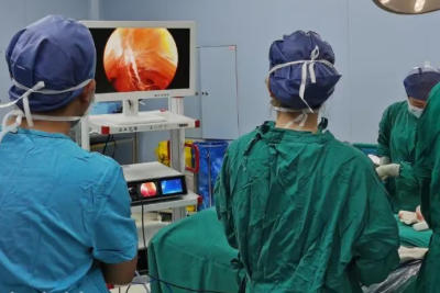
An endoscope consists of an endoscopic camera lens, an LED cold light source, and a screen monitor display. It can be inserted into the body through the mouth or other natural orifices, or through small surgical incisions, extending the surgeon's vision and expanding the surgical field. Simply put, an endoscope is like an electronic eye, allowing visualization of tissues invisible to the naked eye. Familiar instruments like otolaryngoscopes, arthroscopes, and laparoscopes are examples. Endoscopic breast augmentation is a procedure that uses an endoscope to assist in breast augmentation. An incision can be made in the armpit to clearly visualize the internal structure of the breast tissue. After all, "performing surgery under visual guidance" is far safer than "performing surgery blindly."
The surgeon performs the dissection based on the placement of the prosthesis. During the procedure, an electrocautery device is typically used for cavity dissection. This device heats the tissue through high-frequency, high-voltage current generated at the tip of the electrode, achieving tissue separation and coagulation, thus achieving the purpose of cutting and hemostasis. An endoscope allows for easy control of the dissection operation and management of bleeding points. Furthermore, the endoscope provides precise control over the extent of cavity dissection and the placement of the prosthesis.
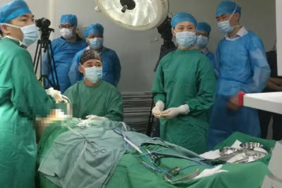
Safe Direct Visualization During Breast Augmentation: Endoscopic breast augmentation overcomes the limitations of traditional "blind" surgery. Under endoscopic imaging, surgeons can perform precise procedures, avoiding damage to blood vessels and tissues. It also significantly aids in cavity separation and implant placement. Key steps such as incision, dissection, electrocoagulation, irrigation, suturing, and repair are all performed under direct visualization, maximizing precision and effectively protecting nerves and surrounding tissues. This results in greater safety during surgery, less postoperative swelling and bruising, and a lower incidence of capsular contracture. The magnification provided by the endoscope allows surgeons to clearly identify nerves, blood vessels, and muscles, eliminating blind spots and minimizing the risk of vascular rupture during the procedure. Even minor bleeding can be detected immediately and stopped with the endoscope.
The surgical incision is small
Since endoscopy is an auxiliary technique, doctors can display the incision on a screen through a smaller incision under visual guidance, making it easier to adjust the position of the implant and the shape of the breast.
Shorter surgery time
With the use of endoscopic technology, doctors can perform surgeries with greater precision, reducing surgical time by 20%-30% compared to traditional methods, and improving the biocompatibility between the body and the implant. Endoscopic technology can also improve flat, sagging, loose, and deflated breasts, resulting in fuller, more prominent breasts.
Faster recovery time
Traditional breast augmentation methods typically require a long recovery period, typically 3-6 months. However, with endoscopic techniques, the recovery time is reduced to one month, allowing patients to quickly return to normal social and work activities.
- Recommended news
- 【General Surgery Laparoscopy】Cholecystectomy
- Surgery Steps of Hysteroscopy for Intrauterine Adhesion
- [Percutaneous Endoscopic Lumbar Discectomy] Percutaneous endoscopic lumbar discectomy for lumbar disc herniation
- [Urology Cystoscopy Section] Cystoscopic Laser Lithotripsy
- [Laparoscopic Urology] Laparoscopic Radical Right Nephrectomy