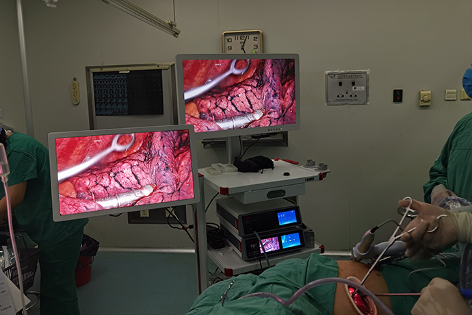
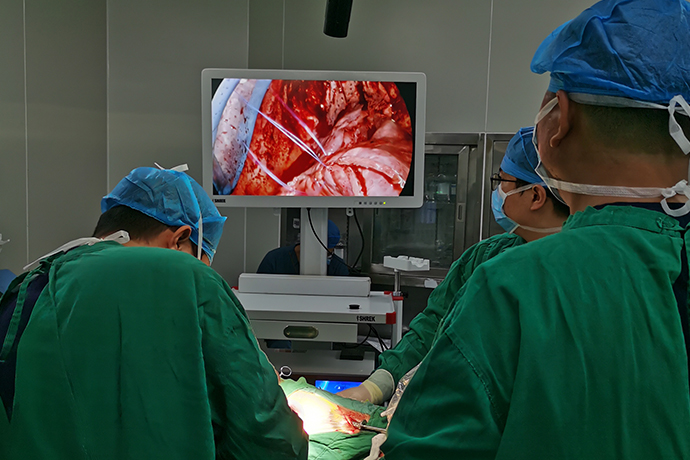
[Thoracoscopic Thoracic Surgery] 4K ultra-high definition thoracoscopic empyema surgery
Release time: 05 Mar 2024 Author:Shrek
What is empyema
The pleural cavity is infected by purulent pathogens and the accumulation of purulent exudate is called purulent pleurisy, or empyema for short.

According to the onset and course of the disease, it is divided into:
(1) Acute empyema: The course of acute empyema generally does not exceed 6 weeks.
(2) Chronic empyema: If the course of the empyema exceeds 6 weeks, it is called chronic empyema.
Main causes
Causes of acute empyema:
A. Pulmonary infection: Pulmonary purulent infection, especially lesions close to the pleura, spreads directly to the pleural cavity. is the main cause of infection.
B. Trauma: open chest injury, lung injury, trachea and esophageal injury, etc. can cause empyema.
C. Spread of adjacent infection focus: mediastinal infection, subphrenic abscess, suppurative pericarditis, etc., can directly erode and perforate the pleura or cause empyema through lymphatic drainage.
D. Hematogenous empyema: In sepsis or sepsis, bacteria reaching the pleural cavity through blood circulation can cause empyema.
E. Thoracic surgery contamination: hemothorax infection, bronchopleural fistula, esophageal anastomotic fistula, etc. occur after surgery.
F. Others: such as spontaneous pneumothorax closed drainage or repeated puncture, secondary infection and rupture of mediastinal teratoma, etc.
Causes of chronic empyema:
A. Improper drainage of acute empyema: The drainage of acute empyema is not timely, the drainage site is inappropriate, the drainage tube is too thin, the insertion depth is inappropriate, or the drainage tube is pulled out prematurely, so that the pus cannot be drained.
B. Foreign bodies: Foreign bodies remain in the pleural cavity, such as shrapnel, cloth scraps, dead bone fragments, etc., which are more common in gunshot wounds and explosion injuries, especially blind tube injuries.
C. Idiopathic infections Infections such as tuberculosis, fungi, and parasites can lead to chronic empyema.
D. Chronic infection of adjacent tissues. Chronic empyema can be caused by infectious diseases such as rib osteomyelitis, subgastric abscess, liver abscess, etc.
Popular groups and symptoms
Predisposed groups: People with infections in the lungs or adjacent tissues of the chest, patients who have undergone chest surgery or operations, and people with low immunity.
Typical symptoms of empyema include fever, chest pain, chest tightness, cough, and sputum production. In addition, patients may also have consumptive symptoms such as weight loss, pale complexion, memory loss, and general weakness. There may be dullness upon percussion at the abscess site and decreased breath sounds on auscultation. . Complications of this disease include pneumothorax, lung atrophy, thoracic invagination, scoliosis, etc.
Surgical options
The purpose of empyema surgery is threefold: to ensure lung recruitment, to remove pus and necrotic tissue, and to remove diseased lung lobes. When formulating treatment plans for patients, the above three core purposes should be comprehensively considered.
Whether to choose endoscopic or open surgery depends on the patient's condition and the surgeon's surgical skills. Detailed medical history questioning and careful CT scan reading are necessary before surgery. Before surgery, it must be determined whether the lung lobes need to be removed at the same time, the degree of pleural thickening and whether there is fiber plate formation.
Generally speaking, stage 1 and stage 2 empyema can be solved through laparoscopic surgery, while open surgery is still recommended for stage 3 chronic empyema. Some professors are using laparoscopic minimally invasive surgery to treat chronic empyema, but it is not mainstream, and it is very popular to promote it. Difficulty.
Due to the conversion of field of view and the application of long instruments, the surgical operation of thoracoscopic surgery is very different from traditional thoracotomy surgery. The surgeon must undergo strict thoracoscopic surgery training in order to master the thoracoscopic surgery operation technology. The large screen and ultra-high-definition camera equipment used in thoracoscopy can enlarge the surgical field of view as needed and clearly display the fine structures. The exposure of the surgical area and the extent of resection are better judged than traditional thoracotomy.
Due to the advantages of mild pain, quick recovery, and clear observation of fine structures, video-assisted thoracoscopic surgery is increasingly used in thoracic surgery and has become the most widely used surgical method in thoracic surgery.
1.Selection of incision
Whether laparoscopic or open, the incision can be made at the 5th or 6th intercostal space. If the upper lobes are well recruited and the pleura is not thickened, the sixth intercostal incision can be more conducive to dealing with underlying adhesions. Because the treatment of empyema is often most difficult near the lower lobe of the lung and the diaphragm.
Of course, the incision of empyema does not need to be rigid. You can also open an operating port at the lowest part of the abscess cavity to treat the lower part, and add another operating port in the third or fourth intercostal space above to treat the upper part.
2. Chest entry (4K ultra-high definition thoracoscopy surgery)
Before surgery, a CT scan was performed to check the degree of pleural thickening and whether there was pus in the chest cavity. Use this to determine what is going on underneath our incision.
If you perform closed chest drainage in advance and discover empyema, leaving a small amount of pus in the chest or frequently flushing with normal saline after the pus is drained will reduce the adhesion of the visceral parietal pleura. This will greatly reduce the difficulty of the operation.
This again involves the issue of the timing of surgery. After the empyema is diagnosed and closed drainage is performed, the earlier the timing of surgery is selected, the better the surgery will be performed. The factors that restrict us from performing surgery as soon as possible include: the original condition does not allow it (such as the acute stage of pneumonia), the patient's physical condition, and other objective factors.
To be fully prepared for possible difficulty in entering the chest, the operating port should be opened first. After reaching the intercostal space, a small retractor should be used to expose the incision, and the intercostal muscles should be gradually incised and continued down to the thickened pleura. We should know the situation below the incision through CT. In short, the pleura is thickened. Incision is a necessary step. Be careful of injuring the lung tissue. You can use your fingers or oval forceps to intermittently add blunt dissection.
After entering the chest, use your fingers to separate it first. If the abscess is not tightly adhered, it can be easily peeled off, clean around the operating port, and then place an incision protective cover. Do not let the incision protective cover trap some pus, because we also need to enter the mirror from the operating port to perform the operation, which will affect the field of view.
Afterwards, the scope is inserted through the operating port and separated using single-port thoracoscopy technology. I am generally accustomed to adding an additional endoscopic hole between the two ribs below the operating hole, and the endoscopic hole can be further expanded to treat more difficult adhesions between the lower lung lobe and the diaphragm. To separate in the direction of the planned endoscopic hole, it is best to clean the parietal pleura there, otherwise the field of view will be greatly affected.
At this point, thoracoscopic empyema evacuation officially begins.
3. Release of the lungs
4. Some details of lung dissection
(1) When freeing, a combination of blunt and sharp methods can be used. Blunt dissection can be performed with a suction device or oval forceps gauze. Whether open or endoscopic, endoscopic vision should be provided because it is safer.
(2) The dissection of the chest top and upper mediastinum must be particularly careful and gentle. Once a large blood vessel is torn, it may be fatal. Important structures such as the subclavian artery and vein, phrenic nerve, and superior vena cava must be noted. It is strongly recommended to operate under the endoscopic view. When the blunt dissection reaches the blood vessel, the speed should be slowed down and the operation should be done carefully under the microscope. The loose area can be bluntly separated with an aspirator. If there is a cord, use an electric hook or ultrasonic to cut it off. Do not tear it blindly. .
(3) When separating lower lobe adhesions, start from the front pericardium, pay attention to protecting the phrenic nerve, and look for the interface between the lower lung and the diaphragm. Always pay attention to whether the diaphragm is damaged. Once damaged, it should be repaired and the interface adjusted in time.
(4) The diaphragm attachment point should be recognized and protected. Generally, caution should be taken when reaching the level of the 9th rib. The diaphragm attachment point cannot be cut off as an adhesion.
5. Peeling of pleural fiberboard
(1) When removing parietal pleural fiberboard, special attention should be paid to preventing damage to extrapleural blood vessels and nerves, such as azygos vein, thoracic duct, esophagus, subclavian artery and vein and mediastinal surface. The phrenic nerve, recurrent laryngeal nerve, superior vena cava and other important structures. If necessary, stripping of important and dangerous locations can be abandoned.
(2) The visceral pleura must be peeled off patiently and carefully. First cut the fiberboard with a blade until normal lung tissue is seen, then use vascular forceps to lift the fiberboard, and use peanuts, scissors, electric knife, knife handle, etc. to peel it off below. When it is difficult to peel off certain areas, the surrounding area can be peeled off as much as possible to leave an "isolated island". If the island still affects lung recruitment, a "well"-shaped incision can be made on the "isolated island". The incision must reach the lung layer pleura.
(3) During the stripping process, for those who have lacerated lung tissue during the stripping process, they should be promptly repaired with small round needles and fine silk threads to avoid omissions.
(4) Ask the anesthetist to inflate the lungs to check where the visceral pleural peeling is not satisfactory and where the lungs are leaking.
(5) Pay attention to detect whether there are tumors in the lungs, whether the lung tissue is damaged or consolidated, etc. If the lung lobes cannot be recruited all the time, then whether to resect the pulmonary lobes together will be evaluated based on the specific circumstances.
(6) When dissociating, it is necessary to thoroughly recognize and separate the pulmonary fissure.
6. Preparation before closing the chest
(1) Flush the chest cavity with large amounts of warm salt water to completely remove pus and necrotic tissue.
(2) To completely stop bleeding, it is best to use an electrocoagulation rod or electric hook or electrosurgery to carefully stop the parietal pleura under the microscope. You can also use hot saline gauze pads to compress and stop bleeding, and use a thoracoscope to carefully explore whether there is active bleeding from top to bottom.
(3) Instruct the lungs to inflate and check the lung recruitment and air leakage again.
(4) Soak the chest cavity in iodophor water for 5-10 minutes. While soaking, clean the incision. For thoracic surgery, iodophor is a good thing. It is less irritating, oily, has good permeability, and iodophor has a sterilizing effect. Therefore, iodophor can be used both as a sterilizer and as an adhesive. For some patients whose residual cavity is not closed and always has pus after surgery, repeated irrigation with iodophor can be used.
(5) A drainage tube is routinely placed in the second rib. For the upper drainage tube, it is recommended to place a fungus-shaped head latex drainage tube, which will cause less damage to the lungs and has a good drainage effect. For pneumothorax patients, especially those whose lungs are covered with bullae, placing a fungiform head drainage tube is the best choice.
(6) Place a 28# drainage tube at the rear and cut a few more side holes.
(7) Decide whether to place a third drainage tube according to the situation.
(8) The removed pus, fiberboard and other tissues should be routinely sent for pathological examination.
7. Postoperative management
(1) The water-sealed bottle should be routinely connected to negative pressure after surgery to promote lung recruitment and adhesion as soon as possible and eliminate dead space. Generally, negative pressure can be connected to the upper chest tube.
(2) Routinely review blood routine, biochemistry, blood gas analysis, and bedside chest X-ray on the morning of the first postoperative day. Find out whether there is anemia, low protein, and whether the lungs have been fully recruited. Depending on the situation, decide whether to transfuse blood or human albumin, etc.
(3) Normal saline can be dripped from the upper chest tube for flushing (generally not needed)
(4) Control the patient’s blood sugar, blood pressure and other indicators after surgery.
(5) Kaiselu can be given routinely on the first day after surgery to stimulate defecation. After the operation, ensure that the patient eats well and has smooth bowel movements.
(6) Adequate nutrition must be ensured, and parenteral nutrition can be added if necessary in the first few days after surgery.
(7) Antibiotics are routinely used after surgery, and the medication can be guided by the bacterial culture results of blood or pus before surgery.
(8) Pay attention to re-examination of chest CT to thoroughly understand the situation of lung recruitment and adhesion, and check whether there is dead space.
Postoperative precautions
Although thoracoscopic surgery is less invasive, it still incises the chest wall and removes part of the lung tissue, which still causes a relatively large impact on the patient's body. Therefore, there are still many things that need to be paid attention to after the operation. Postoperative precautions mainly include the following five aspects:
Strictly quit smoking: long-term smoking will cause patients to increase sputum secretion and weaken sputum discharge function, which can easily cause lung infection. Especially after lung surgery, patients are at a higher risk of lung infection. Therefore, patients with lung surgery are required to start before surgery. Strictly quit smoking for at least 2 weeks, and you need to stop smoking for a long time after surgery.
Eat a light diet and strengthen nutrition: In the early stage after surgery, patients often experience a loss of appetite. At the same time, surgical stress will lead to increased consumption of nutrients. Therefore, the diet should be kept light and easy to digest, eat more protein-rich diet, and supplement nutritional intake to ensure Recovery of the body.
Early mobilization: Both surgery and tumors are factors that significantly increase the risk of thrombosis. Therefore, if there are no contraindications after surgery, patients will be required to move early to prevent the occurrence of thrombosis, and thoracoscopic surgery is no exception. After thoracoscopic surgery, you can usually get out of bed the next day. You can make a gradual transition by sitting up first, sitting next to the bed, then trying to stand, and then walking slowly with the support of family members.
Deep breathing training and coughing and expectoration: Lung tissue contains a large amount of gas under normal conditions. During lung surgery, the lung tissue on the surgical side will collapse. After surgery, the collapsed lung tissue needs to be restored to its pre-operative inflation state, that is, Lung recruitment is also the most important aspect of recovery after lung surgery. This requires patients to take active deep breaths, cough and expel sputum in a timely manner to avoid atelectasis and lung infection and promote the recovery of respiratory function.
The correct way to cough is to breathe abdominally, take a deep breath, hold it and then cough deeply. You can use painkillers appropriately to reduce the pain caused by deep breathing and coughing.
Face postoperative pain: Compared with traditional thoracotomy surgery, thoracoscopic surgery can reduce the pain and shorten the duration, but there will still be a certain degree of pain and it will take several months to recover. The degree and duration of pain vary depending on the It varies from person to person. Therefore, patients maintain a good attitude, believe that the pain will gradually be relieved, and actively cooperate with treatment and rehabilitation activities. If the pain is severe and unbearable, you should tell your doctor promptly and use analgesics appropriately.

- Recommended news
- 【General Surgery Laparoscopy】Cholecystectomy
- Surgery Steps of Hysteroscopy for Intrauterine Adhesion
- [Otolaryngology Nasal Endoscopy] How to Treat Recurrent Rhinitis
- [ENT Surgery: Nasal Endoscopy] Endoscopic Treatment of Nasal Polyps
- [Otolaryngology Nasal Endoscopy] Methods and Precautions for Nasal Bleeding Control under Nasal Endoscopy