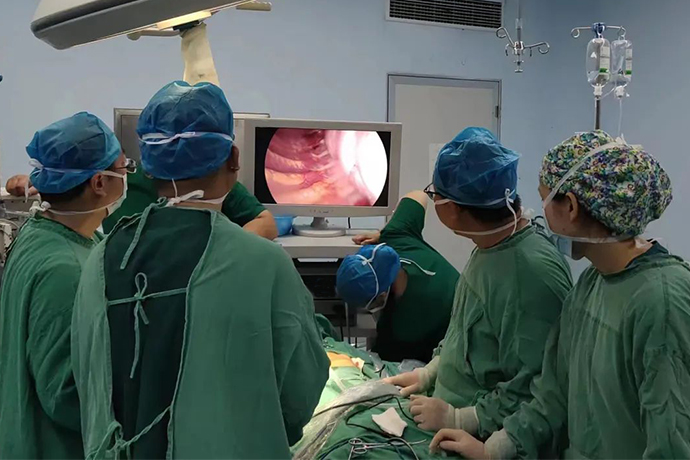
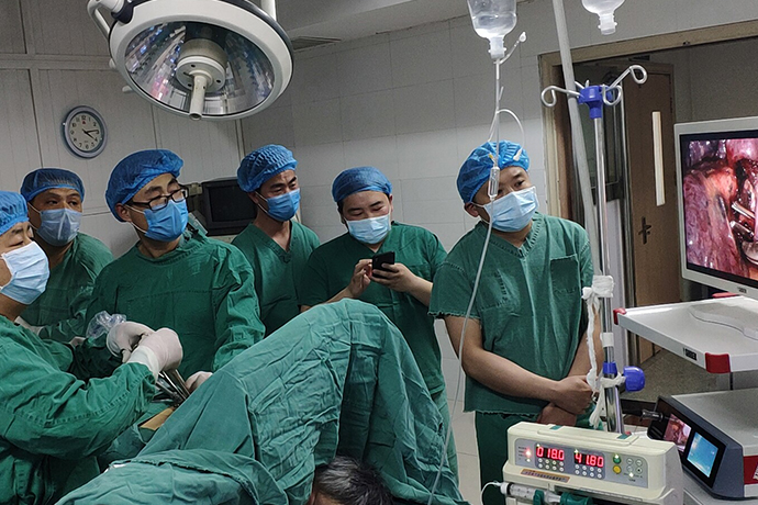
[Thoracoscopic surgery of thoracic surgery] 4K ultra-high definition thoracoscopy for correction of chest wall deformity
Release time: 30 Jan 2024 Author:Shrek
1. What is chest deformity?
Chest wall deformity is generally caused by congenital developmental disorders of the chest wall that cause abnormalities in the shape and anatomy of part of the chest wall. There are not many serious life-threatening cases of this type of lesions. Common lesions include the following: ① pectus excavatum; ② pectus carinatum; ③Poland syndrome; ④Sternal cleft; ⑤Other lesions include thoracic vertebrae-rib deformity (Jarcho-Levin syndrome), rib dysplasia, etc.

Pectus excavatum accounts for more than 90% of all chest wall deformities. Mild cases often show no damage to cardiopulmonary function. However, as the degree of deformity worsens, the shoulders will lean forward, the back is arched, the front chest is sunken, and the abdomen is bulging. Deep inhalation The typical sign of pectus excavatum is the abnormal depression of the sternum. Children with cardiopulmonary compression may develop respiratory and circulatory system symptoms, including palpitations after activity, asthma, precordial pain, reduced vital capacity, and recurrent respiratory infections. In severe cases, it can lead to abnormal cardiopulmonary development and irreversible damage to cardiopulmonary function, which can lead to a sense of inferiority and even a series of psychological problems such as social impairment, autism, and depression.
2. Does chest wall deformity require surgery?
Funnel index = (a×b×c)/(A×B×C), where a is the longitudinal diameter of the infundibulum depression, b is the transverse diameter of the infundibulum, c is the depth of the funnel, A is the length of the sternum, B is the transverse diameter of the thoracic cage, and C is the shortest distance from the sternal angle to the thoracic vertebral body. A funnel index <0.2 indicates mild deformity, a funnel index 0.2-0.3 indicates moderate deformity, and a funnel index >0.3 indicates high degree of deformity.
Surgery is required: If pectus excavatum affects cardiopulmonary function, causes cardiopulmonary burden, or funnel index is >0.2, surgical treatment is required. Therefore, moderate and severe deformities require surgical treatment. For patients whose cardiopulmonary function, body shape, and appearance are affected, early surgical treatment is recommended.
The main treatment methods for pectus excavatum include: sternal lift, sternal suspension and traction, traditional sternal flip surgery-modified sternal flip surgery, and NUSS surgery (new minimally invasive surgery). Single operating port thoracoscopic surgery for pectus excavatum correction (NUSS surgery) uses small incisions on both sides of the chest wall in the front axillary line, and with the assistance of a thoracoscope, a special material orthopedic plate is placed behind the deformed sternum and in front of the heart without cutting off the sternum and ribs. , the surgical trauma is small, the chest wall correction effect is ideal, and the postoperative recovery is fast. 2-3 years after the operation, the orthopedic plate will be removed again according to the patient's chest wall deformity correction status.
Traditional surgical treatment methods involve large trauma, many complications, painful recovery process, and obvious postoperative scars. Currently, the most common surgical method to correct pectus excavatum is Nuss surgery. The surgery is assisted by 4K ultra-high-definition thoracoscopy. A 1.5-2cm incision is made on the chest wall under the armpits on both sides, and the steel plate is carefully inserted from the right side. Enter, pass through the incision on the left side, flip the steel plate to lift up the sunken chest and fix it. The sunken sternum is instantly greatly improved to achieve the purpose of surgical correction of pectus excavatum deformity.
1. Preoperative preparation: Use a soft ruler to measure the distance between the midaxillary lines on both sides of the lowest point of the thoracic depression on the thoracic surface minus 2 cm as the length of the alternative stent. Based on this, select an orthopedic plate that is equal to or slightly shorter than the length of the stent, and use a bender to bend it. The supporting steel plate is shaped and bent into shape. The arc is consistent with the preset lifting height.
2. Marking: tracheal intubation under general anesthesia, supine position, slightly elevated chest and back, and abduction of both upper limbs. Generally, the lowest point of the thoracic cavity or slightly above it is used as the support plate passing plane, and the lowest point of the sternum and two points parallel to it are marked. The highest point of the lateral ribs (where the orthopedic plate penetrates and exits the chest), and the incision (the corresponding intercostal space between the anterior and midaxillary lines).
3. Surgical path: Make a 2 cm transverse incision at the incision mark to separate the subcutaneous tissue and muscles, generally to the highest point of the ribs on both sides. For one-lung ventilation (for example, a single-lumen tube can temporarily stop lung ventilation), a thoracoscope is inserted into the 1st to 2nd intercostal space below the same incision. Under minimally invasive thoracoscopy monitoring, the guide is inserted through the incision into the preselected intercostal space. The edge of the depression penetrates into the chest cavity, closes to the chest wall to separate the retrosternal space, slowly passes through the predetermined support point of the sternum, crosses the mediastinum, and exits at the marked point on the left side of the chest cavity depression edge at the same level.
4. Guide: Connect the support plate to the guide with a thick wire, and guide the convex side of the support plate backward through the retrosternal tunnel. After the support plate is in place, use a turner to turn it 180 degrees to lift the sternum so that the chest wall reaches the desired shape.
5. Fixation: Put the support plate into the fixator, sew the fixator to the rib periosteum, and then sew the fixator, support plate and chest wall muscles together.
6. Suture: After checking that there is no bleeding in the chest, sew several needles with thin silk thread at the entrance of the thoracoscope. Instruct the anesthetist to expand the lungs to remove the thoracic air and then tie the knot. A drainage tube may or may not be placed.
7. Surgical precautions: If the guide passes through the lowest point of the sternum during the operation, if there is no thoracoscopic monitoring, care should be taken to keep it close to the posterior wall of the sternum to avoid damaging the heart. At the same time, ECG monitoring should be closely observed to avoid serious arrhythmia or even cardiac arrest caused by compression of the heart. Fight.

- Recommended news
- 【General Surgery Laparoscopy】Cholecystectomy
- Surgery Steps of Hysteroscopy for Intrauterine Adhesion
- [Otolaryngology Nasal Endoscopy] How to Treat Recurrent Rhinitis
- [ENT Surgery: Nasal Endoscopy] Endoscopic Treatment of Nasal Polyps
- [Otolaryngology Nasal Endoscopy] Methods and Precautions for Nasal Bleeding Control under Nasal Endoscopy