[Gynecological Laparoscopy] Improving surgical skills of 4K laparoscopy
Release time: 12 Sep 2023 Author:Shrek
The difference between laparoscopic surgery and open surgery
1. The abdominal cavity is not incised. It only needs to go through 3-4 small incisions of 0.5-1cm, insert a laparoscopic puncture device and operate it laparoscopically. The abdominal cavity does not need to be exposed to the air.

2. With the help of the camera system, the surgical field of view is fully exposed than in traditional surgery.
3. The parts above the surgical operation will not be interfered by unnecessary operations.
4. Incision, ligation, and hemostasis are mainly completed by electrocoagulation surgery. Foreign bodies at the surgical site are significantly reduced, and pelvic adhesions are reduced.
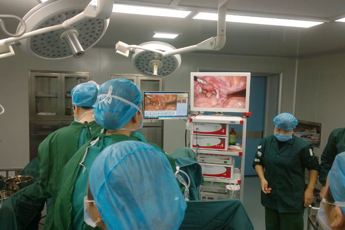
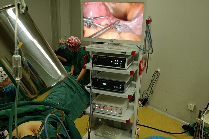
At present, 4K laparoscopic surgery has been widely used in gynecology. Most gynecological diseases and even gynecological tumor surgeries can be performed laparoscopically. In addition to traditional laparoscopy, single-port laparoscopy is increasingly used. Using 4K ultra-high-definition 55-inch monitor imaging to view surgical images, you can see the lesions more clearly, and perform surgery accurately, quickly, and save time. It can not only relieve the patient's pain, but also save operation time, and better assist the doctor as a "third eye".
However, some doctors still say that when it comes to laparoscopic surgery, it is easy to watch others do it, but why do they still find it difficult to do it themselves?
Beginner
Characteristics: I am new to laparoscopy and cannot tell the difference between east, west, north and south after the separation forceps are inserted into the abdomen.
Advanced paths: surgical monographs, simulation boxes, familiarity with the environment and weapons.
1. Monograph on Laparoscopic Surgery
At least one monograph on laparoscopy is required. Understand the history of laparoscopy, master the anatomy related to surgery, and be familiar with related instruments and equipment and their working principles. Practical learning, theory first, books are the best carrier to provide systematic theoretical learning.
2. Simulation box operation
Train hand-eye coordination in the simulation box, such as clamping peas, peeling grape skins, etc.; practice clamping, separation, suturing, and knotting. Suturing and tying knots is the most difficult operation during laparoscopy. If you want to improve quickly, deliberate practice in a simulation box may be the best way.
Teaching hospitals often have endoscopic training centers. Simulation box operation is a compulsory part for graduate students and one of the contents of the final examination for trained physicians.
High-end training can be combined with VR virtual reality, AR augmented reality or even AI artificial intelligence; down-to-earth training can be a variety of simulation boxes purchased from online stores; those with a DIY spirit can even make their own simulation boxes. A cardboard box, a camera, and a laptop can do it, and the training effect is equally amazing.
1. Familiar with the operating room environment and endoscopic equipment
Before surgery, you need to be familiar with the operating room environment and endoscopic equipment.
The environment of the operating room, such as the control of the operating table, how to set up the lithotomy position, how to position the head high and the feet low; such as how to connect the air inlet channels on the wall; the position and connection of negative pressure suction, etc.
Understand the names and functions of each device on the laparoscopic gantry. How to connect optical fiber, how carbon dioxide comes in and out, what do the numbers on the display of the insufficiency machine mean; how are monopolar, bipolar, ultrasonic scalpels and other equipment connected, what are the basic principles; how is the suction device assembled, Trocar How to disassemble, how to install, etc.
These questions are subtle, but important. When you become the chief surgeon, or when you have an emergency ectopic pregnancy in the middle of the night and there are no skilled nurses, you will encounter various problems during the operation, such as the screen is not clear, the air pressure during the operation is not enough, the single and double poles do not work, you have to know the reasons, you have to Know how to solve it.
Primary Stage
Features: Can successfully complete some level 2 gynecological surgeries, such as fallopian tube removal, ovarian cyst removal, etc.
Advanced path: watching peer surgeries, rumination, video review and editing.
1. Watch a fellow surgeon perform surgery
It can be online videos or on-site operations by peers; it can be from this major or from other majors; it can be done well or done horribly like a car accident scene. If you do well, learn from experience; if you do poorly, learn lessons.
In today's era, there are numerous WeChat public account articles, short videos, and surgical videos, as well as unlimited online learning resources, which are a latecomer advantage for young people to learn laparoscopy.
2.Rumination
After the surgery, on the way home or on the subway, reflect on what went well during today's surgery, what needs to be improved, where the problems are, and how to improve. Detailed rumination can even exceed the training effect of hands-on operations. Only by constant thinking every step of the way can the surgery be done better and better!
3. Record, review and edit videos
Recording surgical videos has several benefits:
Watch and learn by yourself
Teaching, peer exchange
If postoperative complications or even doctor-patient disputes occur, you can look for evidence
Looking back at the surgery video is the most detailed rumination. Find out the problem by watching your own surgery video: Why does bleeding occur during this step? Are there too many useless moves here? Can it be simplified? Is there anything wrong with exposure?
Look at your own surgery from the perspective of a bystander and constantly improve. Watching back the video is also a process of learning anatomy. Comprehension of anatomy is the basis for improving surgical skills.
In addition, editing videos is also a very effective way to improve your skills. Whether it is for retention, teaching, video competition, or conference communication, editing videos is a process of eliminating the unnecessary and retaining the essential, and it is also a process of learning and improvement. Videos can be edited to highlight anatomical structures, refine the key points of surgical operations, and eliminate wasteful movements. This process is even better than training on the operating table.
Intermediate and advanced stage
Features: Congratulations, you are now able to skillfully complete level 3 to 4 laparoscopic surgeries!
Advanced path: Same as before. Focus on reviewing your own surgery.
This stage is no longer limited to completing an operation, but needs to achieve the following goals: the operation will be smoother, shorter, less bleeding, clearer anatomy, fewer complications, and faster patient recovery.
At this time, while learning from others, you need to carefully consider your own surgery. After years of surgical operations, you will definitely develop your own surgical style, discover your own strengths, and understand yourself more clearly. This is not only a way to improve surgical skills, but also requires this kind of mentality to grow in life.
Of course, the rumination mentioned above, deliberate training in the simulation box, and watching other people's surgeries also apply at this stage.
Master level
Characteristics: Expert in the industry, protagonist of surgical demonstrations, being imitated and sought after.
Advanced path: competition among masters.
An important way to practice deliberately is to break down a big goal into small details, divide the content to be trained into targeted small pieces, and practice each small piece repeatedly.
For example, laparoscopic training can use this method. Compared with traditional open surgery, the biggest change in laparoscopic surgery lies in the operating instruments, such as aspirators, separation forceps, ultrasonic scalpels, needle holders, etc.
Laparoscopic skills can be improved through daily surgical operations. But practice on stage alone is not enough. If you want to master every instrument quickly, you need more training off the stage.
Training for each instrument can be carried out through deliberate practice. Deconstruct the key points of operation of each instrument, extract the core movements, and practice them repeatedly to achieve mastery and "the sword and man are one". Below, we break it down one by one.
Deliberate practice of attractors
The suction device has many functions. In the past, Dr. Ding Jin summarized the seven major functions ��� Do you always get scolded by the director when you are an assistant? You need to know these techniques, but the core moves are sucking and punching. Implemented on the hand, it means pushing forward and pulling back with the thumb (when operating with small hands, it may be pushing forward with the thumb + pulling back with the index finger).
The deliberate training for the attractor mainly involves practicing the two movements of pushing and pulling. There is no need to waste precious time on the operating table, and there is no need to sit in front of the simulator and hold the suction device directly to complete the deliberate practice of the suction device.
Repeat these two movements of pushing and pulling, increase the frequency, train the thumb and related muscle groups, and implant the memory of these movements into the brain. After deliberate practice, your muscles and brain will change accordingly. This process of deconstruction and practice may seem boring, but it is extremely effective.
Deliberate practice of separating forceps
Likewise, the core actions of a separator plier are separation and clamping. Hold the separator pliers directly and, looking straight down, pinch pieces of paper, peas, millet, or anything else within your reach. Practice to clamp harder, faster and more accurately. The left and right hands alternate or work together to complete the deliberate practice of separating the pliers.
In the open era before laparoscopy, surgeons mostly used hemostats for surgical procedures. Being able to accurately clamp blood vessels to stop bleeding is an important basic skill. Mr. Qiu Fazu, a leading figure in the surgical field, once sprinkled a handful of rice on the bed when he was practicing, and then used hemostatic forceps to pick up the rice one by one. This was also deliberate training.
Deliberate practice with ultrasonic scalpel
The core of ultrasonic scalpel action is precision. The difference between master surgeons and ordinary surgeons in the use of ultrasonic scalpel lies in the word "precision". Again, there is no need to mobilize an army, just a discarded ultrasound scalpel can be used to practice.
For example, crumple a piece of A4 paper into a ball, and use the blade of a knife to hold each wrinkle while looking directly at it. When clamping different wrinkles, change the direction of the cutter head to ensure that the cutter head is perpendicular to the wrinkles. Feel the different clamping strengths and experience the movement between the two sides of the cutter head... making clamping faster and more accurate.
Deliberate practice of suturing techniques
Suturing is the most difficult technique to master in laparoscopic surgery. This is inseparable from the skillful use of the needle holder. So how do we carry out deliberate practice of suturing techniques? The core movements of suturing include holding the needle, advancing the needle, and removing the needle.
The simplest form of deliberate practice requires only a needle holder and a needle. Try to find a piece of paper, a layer of cloth or an orange peel, and practice holding, inserting and removing the needle under direct vision. Practice with full concentration and let the repeated movements reshape the muscles and change the brain. You will definitely be much more skilled the next time you suture on the stage.
Deliberate practice of knotting techniques
The core part of knotting technology: the winding between the needle holder and the separation forceps.
The equipment for practice can be a needle holder, a pair of separation forceps, and a suture, or two separation forceps and a thread. You can even practice anywhere by holding two long sticks with their heads wrapped around each other...
However, the most fearful word in everything is seriousness. If you hold a long stick in each hand and practice laparoscopic knotting techniques while walking, is it possible that you will not be able to learn laparoscopic techniques?
Common misunderstandings
1. Laparoscopic surgery = minimally invasive surgery
"Many people think that laparoscopic surgery = minimally invasive surgery. But in fact, laparoscopic surgery ≠ minimally invasive surgery." Laparoscopic surgery, like traditional laparotomy and transvaginal surgery, is just a choice of surgical route. . Minimally invasive surgery is a surgical concept. As long as the surgical method, patient's physical condition, and indications are the most suitable, and the surgery can bring maximum benefits to the patient, it is in line with the concept of minimally invasive surgery.
During the surgical operation, for the resection of lesions, the laparoscopic operation is the same as the traditional open surgery, and standard surgical steps need to be followed. Laparoscopic surgery conforms to the minimally invasive concept, but it may also cause "major trauma". Like traditional laparotomy, it also has the risk of damaging surrounding important organs, especially when the scope of the surgical operation is combined with severe dense adhesions, such as large blood vessel damage, Ureteral injury, intestinal injury, etc.
In addition, laparoscopic technology itself will bring special complications, such as CO2 embolism, subcutaneous emphysema, etc. The overall complication rate of laparoscopic technique is 2.58%~3.33%, of which the incidence rate of organ and blood vessel injury is 0.32%~0.37%. Moreover, during laparoscopic surgery, if complications occur that are difficult to handle under the microscope, the patient needs to be converted to traditional laparotomy.
2. Laparoscopic surgery is not as clear as laparotomy surgery
Some people are also worried that the operating field of view of laparoscopic surgery is limited. Will it be less clear than laparotomy surgery? This is also a misunderstanding.
Laparoscopic surgery uses a special light conduction imaging system to project the surgical site on a screen through a special image display system. The surgeon performs the surgery while looking at the screen like watching a movie.
"With the advancement of technology, currently commonly used projection lenses have a magnifying effect, and through high-end image processing systems, the images on the screen are often displayed in 4K ultra-high definition." Therefore, in essence, the anatomy seen under laparoscopy is much smaller than the Traditional laparotomy allows for more precise visual inspection, and with the help of the advantages of instruments, surgical operations are also more precise.
3. Laparoscopic surgery will not be “clean” enough
"Some patients are also worried that laparoscopic surgery will not be done cleanly." This kind of worry is unnecessary. First of all, laparoscopic surgery also follows strict surgical procedures, and the scope of surgical resection is no different from traditional laparotomy. However, when performing myomectomy for multiple uterine fibroids, whether it is traditional open surgery or laparoscopic surgery, there is a possibility that the fibroids that are larger in number, deeper in growth location, and smaller may not be completely removed.
Of course, compared with traditional abdominal myomectomy, laparoscopic myomectomy may indeed cause a lack of direct touch of the lesion. After myomectomy for multiple uterine fibroids, there is a possibility of recurrence regardless of the surgical method.
4. Postoperative abdominal bloating is caused by the lack of emptying of gas in the body
Patients will ask after just completing the operation: "My stomach is very bloated. Did the air that was injected be removed at the end of the operation?" In this regard, she explained that at present, the vast majority of laparoscopic surgeries are performed by injecting gas into the abdominal cavity. Inflating CO2 to expand the abdominal cavity provides better space for surgical operations and improves surgical safety. However, the abdominal cavity must be evacuated at the end of the operation.
"There are many reasons for postoperative abdominal distension, mainly due to incomplete recovery of gastrointestinal function after surgery and intestinal flatulence. Usually, most of the flatulence can be relieved after anal exhaust." Therefore, after laparoscopic surgery, there is usually Patients are encouraged to get up early and take appropriate activities to promote the recovery of gastrointestinal function.
5. Cancer is not suitable for laparoscopic surgery
Many people believe that cancer is not suitable for laparoscopic surgery. In fact, currently, laparoscopic technology can be applied to almost all benign tumors in gynecology, such as uterine fibroid removal, ovarian benign teratoma removal, ovarian endometriosis cyst removal, etc.
The indications of laparoscopic surgery for gynecological malignant tumors are also constantly expanding. Evidence-based medical evidence shows that there is no difference in tumor outcomes between laparoscopic surgery and traditional laparotomy for early-stage endometrial cancer. There are also retrospective studies showing that laparoscopic radical resection for early-stage cervical cancer is also safe and effective. Increasing evidence demonstrates the safety and effectiveness of laparoscopic technology in the field of gynecological oncology.

- Recommended news
- 【General Surgery Laparoscopy】Cholecystectomy
- Surgery Steps of Hysteroscopy for Intrauterine Adhesion
- [Otolaryngology Nasal Endoscopy] How to Treat Recurrent Rhinitis
- [ENT Surgery: Nasal Endoscopy] Endoscopic Treatment of Nasal Polyps
- [Otolaryngology Nasal Endoscopy] Methods and Precautions for Nasal Bleeding Control under Nasal Endoscopy