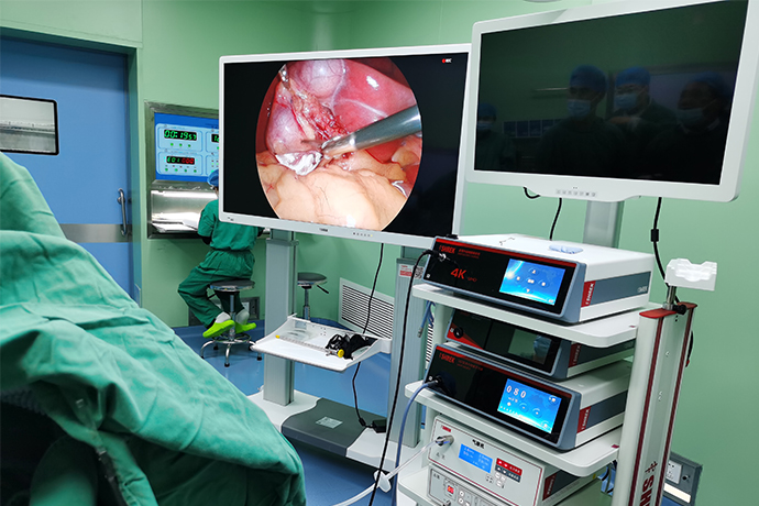
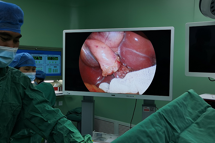
[4K Basics] 4K Ultra HD Endoscope Camera System Technology
Release time: 09 Mar 2022 Author:Shrek
Generally, 4K ultra-high-definition image quality is mainly reflected in five aspects: resolution, color depth, color gamut, contrast ratio and frame number.

Resolution: It is 4K, that is, 3840×2160 pixels, which is four times that of traditional high-definition 1920×1080. The more pixels a monitor can display, the finer the picture, and the more information can be displayed in the same screen area, so resolution is one of the most important performance indicators.
Color depth: The color depth specified in the Ultra HD Blu-ray standard is 10bit, that is, 1.07 billion colors can be displayed (each color in RGB has 2 to the 10th power, which is 1024 levels, and the RGB three colors have a total of 1.07 billion (1024). ×1024×1024) color matching). The 10-bit effect has smooth color transition, rich and full colors, while the ordinary Blu-ray standard supports 8-bit color depth, with only 16.77 million colors, and the gap is huge.
Color gamut: BT.2020 color gamut standard. The BT.2020 color space has been positioned by the ITU International Telecommunication Union as an image signal color gamut standard in the 4K/8K era, and it is also one of the ultra-high-definition Blu-ray standards. The common Blu-ray standard uses the BT.709 color space, which covers only 35.9% of the BT.2020 color space, which is currently the largest color space in display devices, covering 75.8% of the CIE 1931 color space.
Contrast: HDR high dynamic range images. HDR is not the first concept proposed by Ultra HD Blu-ray, but it is crucial to the improvement of picture quality, and it is also the most striking place in the Ultra HD Blu-ray standard. HDR, high dynamic contrast ratio, can take care of dark parts and highlight details on the screen at the same time, bringing a dynamic range closer to human vision. Ordinary TVs, projectors and other display devices have limited contrast performance due to the brightness limit, that is, the SDR (Standard Dynamic Range) level. Generally, the contrast ratio of the LCD screen of our mobile phone is only 1000:1. With the introduction of HDR, a wider 100,000:1 contrast ratio can be achieved.
Frame rate: 60p. The number of frames represents the number of pictures contained in a 1-second video. The higher the number of frames, the smoother the picture observed by the human eye will be, and there will be no intermittent feeling.
4K Laparoscopic Technology
In 2004, the Digital Cinema Initiative (DCI), which is composed of seven major Hollywood film companies, revised and launched its technical document 4.0 industry standard, which stipulates that the definition of digital cinema is divided into two levels, of which the higher level is DCI. The amount of information in 4K (4 096×2 160 resolution, 24 frames/s) is more than 4 times that of HDTV.
Currently, ultra-high-definition laparoscopes with 4K resolution have enabled surgeons to see anatomical structures with greater realism and depth due to their superior image detail, spatial perception, and color accuracy.
In 2018, 4K ultra-high-definition laparoscopy has entered the domestic market. Based on some data collected in this article, it is found that many physicians have the following experience: Under the field of Subtle differences are reflected, tiny blood vessels, tiny nerves, and lymph nodes that are usually indistinguishable from surrounding adipose tissue are clearly discernible under 4K ultra-high-definition laparoscopy, ensuring that the precise operation of lymph node dissection and neurovascular protection can be truly realized. Finding and maintaining is also more precise and convenient.
The 4K laparoscopic system has important research value and application prospects. At present, various fields such as simulated medicine, cardiac surgery, and general surgery have begun to gradually incorporate 4K laparoscopic equipment, and more and more fields have begun to accept 4K laparoscopic systems.
Indications
In the field of general surgery, including liver, gallbladder, pancreas, spleen, stomach, colorectum, appendix, hernia, thyroid and other subspecialties, the 4K laparoscopic system can be used as an optional laparoscopic surgery platform. In view of the fact that the 4K laparoscopic system can provide a higher-definition surgical field and finer detail resolution, it has more prominent advantages in the grasp of membranous anatomy, the identification of fine blood vessels or nerves, and the identification of the boundaries of lymph node dissection. Therefore, its application in gastric, colorectal, pancreas, thyroid, weight loss, hernia and other operations is more practical.
4K Laparoscopic Technology
1.1 Technical basis The basic surgical technology of 4K laparoscopy is based on the basic surgical technology of ordinary high-definition laparoscopy. The participating surgeons should undergo professional training in the laparoscopy training center, be proficient in various basic operation skills of laparoscopy, be familiar with the anatomical structure of the abdominal cavity, and be proficient in the manipulation and use of 4K laparoscopic lenses.
1.2 Visual field exposure and grasp The 4K laparoscopic system is similar to the traditional laparoscope. According to the angle of view of the lens, it can be divided into 0° scope and 30° scope. Currently, the 30° scope is the most commonly used in the field of general surgery. The integrated camera system and autofocus system are used in the 4K laparoscopic system. The endoscope arm can change the viewing angle through the knob on the camera base to adjust the surgical field of view. At the same time, after auto-focusing, the focal length can be fine-tuned through the button according to the needs of the operation, which can effectively reduce the shaking of the surgical field of view, thereby improving the visual experience of the operator and reducing the difficulty of operating the endoscope.
1.3 Screen imaging distance In order to maximize the advantages of 4K high-definition imaging technology, compared with traditional high-definition monitors, the monitors used in the 4K laparoscopic system are larger in size, 31-inch or 55-inch (16:9) monitors. Since the screen size has changed from before, the optimal viewing distance between the surgeon and the screen has also changed. Currently, 4K laparoscopic systems recommend using a monitor no smaller than 55 inches to achieve the best visual effect. It is recommended that the screen be placed at a distance of 150-200 cm from the surgeon to perform surgery to reduce eye fatigue of the surgeon.
1.4.1 Technical Features It can provide the surgeon with a clearer surgical field of vision and vivid images, and the significantly enhanced realism and sufficient magnification can bring better positioning to the surgeon, thereby improving the precision of surgery. Delicate anatomical imaging can improve the operator's anatomical identification, so that the fine anatomy can be completed more smoothly. Therefore, the 4K laparoscopic system is more recognizable than the traditional high-definition laparoscopic system, and has a lower probability of operating errors, which can help physicians to easily identify the relationship between important anatomical structures and surrounding tissues. In addition, the magnification advantage of 4K laparoscopy can relieve eye fatigue compared with traditional high-definition laparoscopy.
1.4.2 Advantages in different surgeries
1.4.2.1 Stomach surgery The main advantage of the 4K laparoscopic system in gastric cancer surgery is that the recognition of various anatomical levels and blood vessels under laparoscopy is greatly enhanced. When performing subpyloric lymph node dissection, the identification of the fusion of the mesogastric and transverse mesocolon can be improved, so that the separation of layers can be more accurate, and the damage of the mesocolic blood vessels can affect the blood supply of the colon. The superficial anterior pancreaticoduodenal vein branches can be more clearly identified and predicted, and unnecessary damage to these branches can be avoided when dealing with the right gastroepiploic vein; when dealing with the root of the right gastroepiploic artery and dissection of lymph nodes , the distinction between pancreatic tissue and lymphatic adipose tissue is more clear and accurate, which can help to avoid accidental damage to the pancreas; during lymph node dissection, the vagus nerve branch on the surface of the common hepatic artery can be fully identified and protected; during lymph node dissection, for The Gerota fascia on the upper edge of the pancreas, that is, the posterior boundary of this group of lymph nodes, can be more clearly identified, so that the dissection of this group of lymph nodes can meet more standard boundary requirements; the 4K magnification effect can make the boundary clearer and avoid Damage to the portal vein caused by misjudgment of anatomical tissue. In addition, the 4K laparoscopic system has richer color levels in the surgical field of view, which can help distinguish the subtle differences between pancreatic tissue and adipose tissue.
1.4.2.2 Colon surgery Since the rise of the concept of complete mesocolic resection, one of the anatomical focuses in colon cancer surgery is the identification and separation of the membranous structure and the complete anatomy of the mesentery. The 4K laparoscopic system can more clearly present the boundary line between the membrane and the membrane with the characteristics of "yellow and white junction"; through the clear identification of the direction of the microvessels on the membrane surface, the ability to identify the membrane can be further strengthened, enabling the operator to perform more accurate measurements. Complete mesangial excision. Under the 4K high-definition laparoscopic system, it is easier for surgeons to identify structures such as the mesocolon, Toldts gap, Gerotas gap, and anterior pancreaticoduodenal fascia, which can better complete the complete mesentery resection while protecting the pancreaticoduodenal fascia. Important anatomical structures such as the denum, reproductive vessels, ureter, etc., reduce the risk of surgery, improve the safety of surgery and radical oncology.
1.4.2.3 Rectal surgery The completion of total mesorectal excision during radical rectal tumor resection is critical to the patient’s postoperative outcome. In the process of finding and maintaining the plane, the microvascular structure between different anatomical layers is more clearly defined. Therefore, it provides a more accurate and objective visual basis to guide the identification, search and maintenance of the correct anatomical level. In addition, easily damaged nerve structures such as the epigastric plexus, hypogastric nerve, pelvic plexus, and neurovascular bundles can be displayed more clearly during the anatomical process of the 4K field of view, which is helpful for more accurate intraoperative protect these nerves. Compared with the traditional 2D laparoscopy system, the 4K laparoscopic system can more clearly display the structure of the narrow pelvic floor space, and the observation of the anatomical structures such as the location of the end point of the mesorectum, the levator hiatus, and the internal and external anal sphincter space is more accurate. Contributes to the precise anatomy of sphincter-preserving surgery for ultra-low rectal cancer.
1.4.2.4 Pancreatic surgery Pancreatic surgery is a difficult point in laparoscopic surgery due to the numerous branch vessels that need to be severed, the complex surrounding anatomical structures, and the variety of postoperative reconstruction methods. 4K laparoscopy improves clarity and relieves some of the difficulties. Under the 4K laparoscopic system, the surgeon can not only accurately identify the anatomy of the gastric and colon related membranes around the pancreas, identify the fatty tissue of the lymph nodes and perform accurate dissection, but also more clearly identify the portal vein, the spleen blood vessels and the small and small surrounding pancreatic glands. Branches, the operator can accurately complete the dissection and ligation of each branch blood vessel, reducing the risk of bleeding. In addition, under the 4K laparoscopic system, the surgeon can clearly see the pancreas, pancreatic duct, bile duct and other structures, thus ensuring high-quality digestive tract reconstruction. In addition, due to the remarkable magnification effect of 4K laparoscopy, it can improve the operator's sense of visual distance during the fine operation of sutures and knots in pancreatic surgery, which can reach or be close to the effect of 3D laparoscopy.
1.4.2.5 Hernia surgery (1) Inguinal hernia: The anatomy of the inguinal area is a difficult point in the operation. There are spermatic cords, round ligaments, reproductive blood vessels, etc. passing through the hernia sac. If the hernia sac is congenital or has been onset for a long time, the hernia sac and sac The relationship between the inner tissue is dense and the dissection is difficult, so the operator's identification of the anatomy is particularly important. Under the 4K laparoscopic camera display system, the relationship between the peritoneum, preperitoneal fascia, transverse abdominal fascia, and preperitoneal fat is clearer, which can guide the operator to avoid entering the wrong level, which is conducive to the stripping of the hernia sac; stripping the hernia sac It is easier to identify the adjacent anatomical structures such as spermatic vessels, vas deferens, etc., and the surgeon can accurately separate the hernia sac to avoid damage to the surrounding structures. At the same time, the anatomical identification of regions such as the crown of death and the danger triangle is clearer, which enhances the safety of the operation. (2) Incisional hernia or parastomal hernia: The magnification and high-definition field of view of the 4K laparoscopic system can more accurately identify the level of the intestinal adhesion tissue, making the separation of intestinal adhesions safer. (3) Hiatal hernia: It helps to identify the hernia sac and pleura in the mediastinum; it helps to avoid accidental injury to the deep abdominal aorta when suturing the angle of the diaphragm; it helps to protect the vagus nerve.
1.4.2.6 Thyroid surgery The advantages of the 4K laparoscopic system for thyroid surgery are mainly reflected in: the color of the parathyroid gland is often similar to that of fat or part of the thyroid tissue, which is easy to cause accidental injury. Under the higher color resolution of the 4K laparoscopic system, the identification of the parathyroid gland can be more accurate, which is helpful for the protection of the gland; the fine blood vessels of the parathyroid gland can be identified in the 4K field of view, thereby protecting the The tiny blood vessels of the parathyroid gland; when protecting the thymus tissue, precise membrane dissection operations are required, and the capsule thickens significantly under the 4K laparoscopic view, which is more advantageous for the identification of membrane dissection. In addition, in the 4K field of view, the tiny blood vessels on the dorsal side of the thyroid near the recurrent laryngeal nerve and the anatomical structure where the recurrent laryngeal nerve enters the larynx are displayed more clearly, which can avoid the bleeding of the blood vessels and lead to the contamination of the surgical level and the surgical field of view. , causing erroneous damage to the nerves. At the same time, the delicate resolution of 4K laparoscopy is also of great significance for neuroprotection during lymph node dissection.
1.4.2.7 Bariatric surgery In gastric bypass surgery, the operation under the 4K laparoscopic system can help to clearly identify the avascular plane and achieve avascular incision; the establishment of the retrogastric tunnel makes it easier to cut off the retrogastric blood vessels. Accurate, in the presence of a large amount of adipose tissue, the ability to identify the HIS angle is strengthened to complete the construction of the gastric bag; it is beneficial to complete the gastrojejunal anastomosis in a small space, and has more advantages for laparoscopic suture, easy to adjust and hold The direction between the needle device and the suture needle facilitates the transfer of the suture needle between the instruments.
1.4.2.8 Liver surgery High-resolution images provided by 4K laparoscopy can be more helpful in identifying liver surface lesions. With higher color discrimination, it can help to more accurately delineate and determine the anatomical resection plane. In addition, in the process of resection, the tissue structure can be clearly distinguished, which is conducive to the identification of bile ducts and blood vessels at all levels in the liver section, so that the corresponding blood vessels and bile ducts can be clamped and cut off more accurately.
Advantages of 4K Laparoscopy
The 4K laparoscopic system can provide better resolution (2160 x 3840), ultra-high-definition 50 frame rate, viewing angle operation coordination, visual acuity, color resolution, noise and flicker suppression, and traditional ordinary 2D endoscopes Compared with the camera system system, it can bring a better surgical field to the surgeon, and the position of the organ and the context of the surrounding tissue can be displayed more clearly. At the same time, the bleeding rate in the hands and the incidence of postoperative complications are greatly reduced.

- Recommended news
- 【General Surgery Laparoscopy】Cholecystectomy
- Surgery Steps of Hysteroscopy for Intrauterine Adhesion
- [Laryngoscopy in Otolaryngology] Tonsillectomy
- [Otolaryngology Otoscopic Section] Excision of Cholesteatoma in the External Auditory Canal
- [Otolaryngology Nasal Endoscopy] How to Treat Recurrent Rhinitis