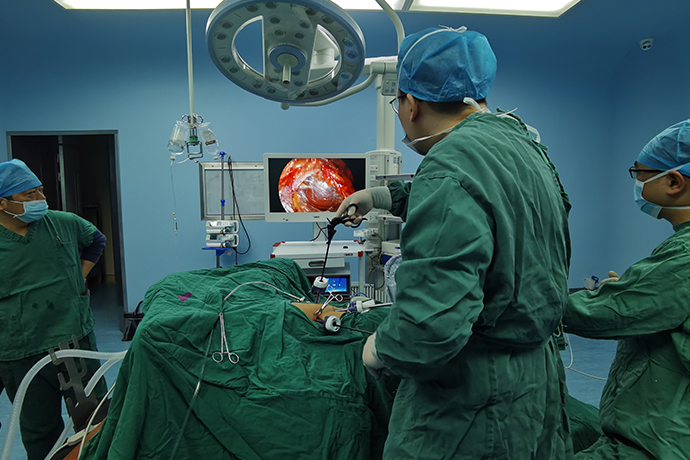
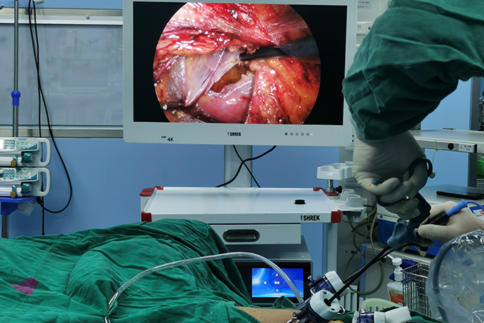
【General Surgery Laparoscopy】Liver Tumor Resection
Release time: 25 Jan 2022 Author:Shrek
Laparoscopic hepatectomy is the mainstream of the development of liver surgery today. Compared with open hepatectomy, laparoscopic hepatectomy has many advantages, including enlarged surgical field, clearer intraoperative tissue structure display, less bleeding, and postoperative incision. Less pain, faster recovery of organ function, less abdominal adhesions, less abdominal scars, shorter hospital stay, and less impact on work and life after discharge.

Anesthesia and preoperative preparation
1. Anesthesia
General anesthesia with endotracheal intubation is generally selected.
2. Preoperative preparation
(1) Liver function assessment: Liver function must be comprehensively assessed before liver resection.
(2) Examination of the function of important organs: The functions of important organs such as the heart, lungs, and kidneys are judged to be able to tolerate liver resection.
(3) Correct anemia and hypoproteinemia.
(4) Perform an enema on the night before the operation and on the morning of the operation day, or take an oral laxative for bowel preparation the day before the operation.
(5) Stomach and urinary catheters were inserted on the same day.
(6) Prophylactic use of antibiotics.
Indications
1. Limited to marginal, superficial, less severe adhesions, and not too large tumors, especially marginal liver lesions located in the left lateral lobe and the anterior segment of the right liver.
2. The tumor capsule is intact, excluding intrahepatic metastasis, portal vein tumor thrombus, hepatic portal lymph node enlargement and distant organ metastasis.
3. Liver function is good, no ascites, jaundice, heart, lung, kidney and other important organ functions are basically normal.
4. The size of the tumor should not exceed 7-10 cm, and it is difficult to operate if the tumor is too large. Hemangioma does not exceed 15cm.
Surgical Steps
General steps of surgery:
1. Establish pneumoperitoneum and explore
The upper edge of the umbilical ring was incised about 1 cm to establish pneumoperitoneum. Explore the abdominal cavity with a laparoscope. If laparoscopic surgery is possible, insert a trocar (Trocar) in the corresponding position.
Trocar: Normally 5
Observation hole: about 2cm above the right side of the umbilicus. Main operation hole: 1-2cm at the lower border of the liver on the median line, about 2cm at the lower border of the liver on the right midclavicular line, and an additional hole at the bottom of the gallbladder if necessary
Auxiliary operation hole: medial to the midclavicular line below the left costal margin, 1 cm below the costal margin of the right anterior axillary line
2. Tumor resection
① Mark the liver resection line;
②Separate and cut the hepatic ligament;
③ Take out the tumor.
3. Treatment of liver wounds
If there is no obvious bleeding and bile leakage on the liver section, no further treatment is required; if there is active bleeding on the wound surface, try to stop the bleeding, and suture, electrocoagulation and other methods can be used. Finally, the pedicled greater omentum was applied to the liver section.
Right hepatic blood flow occlusion technique
In the order of first → third → second porta hepatis
Steps: dissect the hepatic porta; dissociate the liver; dissect the liver parenchyma
free
hepatic round ligament
falciform ligament
Left and right coronary ligaments (left and right inferior phrenic arteries) left and right deltoid ligaments
Adjacent ligaments: liver and stomach, liver and colon, liver and duodenum (prepare for hepatic portal block), liver and kidney
Due to the anatomical relationship between the left hepatic vein and the middle hepatic vein, freeing the second hepatic porta is not overly required.
Cut and ligate the short hepatic veins one by one from bottom to top
Provoking left and right half liver to compress and stop bleeding
Separating the clamp to control the vascular hemorrhage
CUSA/Ultrasonic Bare Pipe, Branch Treatment
CUSA naked blood vessels, ligation nails/titanium clips to clamp blood vessels, ultrasonic scalpel to cut off the liver, bipolar wound hemostasis
auxiliary small incision
segmented liver
inventory
Place the drainage tube
Check content:
Whether there is active bleeding, whether there is bile leakage, whether the ligature is loose
Sutures are often required if abnormal bleeding occurs

- Recommended news
- 【General Surgery Laparoscopy】Cholecystectomy
- Surgery Steps of Hysteroscopy for Intrauterine Adhesion
- [Laryngoscopy in Otolaryngology] Tonsillectomy
- [Otolaryngology Otoscopic Section] Excision of Cholesteatoma in the External Auditory Canal
- [Otolaryngology Nasal Endoscopy] How to Treat Recurrent Rhinitis