【Hysteroscopy】Submucosal Myoma Hysteroscopic Resection
Release time: 13 Apr 2021 Author:Shrek
Classification
• Histology
Leiomyomas (also called fibroids) are tumors in which blood vessels made up of smooth muscle fibers surrounded by collagen fibers do not coincide. They are firm and can be calcified. These benign tumors must be distinguished from leiomyosarcoma.
1. The round ligament
2. Fallopian tube
3. The fundus of the uterus
4. Proper ovarian ligament
5. Uterine cavity
6. Endometrium
7. Myometrium
8. Broad ligament mesentery
9. Uterine artery
10. The ureter
11. Cervical canal
12. Uterosacral ligament
13. The outer mouth of the uterus
14. Vagina
• Leiomyoma
Depending on the location of the leiomyoma relative to the largest transverse diameter of the myometrium, three types of leiomyoma can be distinguished:
1. Uterine leiomyoma, located on the surface of the uterus on the peritoneal side;
2. Intermural lymphoma, embedded in the muscle wall of the uterus;
3. Submucosal fibroids, protruding into the uterine cavity;
4. Only submucosal fibroids can be treated by surgical hysteroscopy.
• Submucosal fibroids
According to the degree of intramural development, three levels of submucosal fibroids can be identified:
1. Grade 0: Development is limited to the uterine cavity (with a pedicle or implanted base);
2. Grade 1: Partial intramural development (internal cavity composition> 50%);
Level 3 and 2: Mainly intramural development (lumen<50%).
Indications
-Symptomatic leiomyoma: uterine bleeding, postmenopausal onset, dysmenorrhea, infertility, history of early miscarriage;
-Submucosal positioning (grade 0 and 1; grade 2, if the thickness of the lateral myometrium of the leiomyoma is> 5mm);
-Size <50 mm;
-Single leiomyoma or bifocal positioning, if the size is small.
Contraindications
-Contraindications to anesthesia;
-Genital infections;
-Pregnancy;
-Multiple leiomyomas (indications for open multiple myoma resection).
Preoperative period
• Ultrasonography
Pelvic ultrasound through the abdomen and intravaginal route depicts the size and grade (S) of submucosal fibroids. For grade 2 submucosal fibroids, ultrasound is used to examine the distance between the outer edge of the leiomyoma and the serous membrane of the uterus to assess the risk of intraoperative uterine perforation. Ultrasound is also used to search for related subcutaneous or intramural fibroids.
Saline versus hysteroscopy can also be performed, which involves injecting saline into the uterine cavity during successive scans.
• Diagnostic hysteroscopy
If the ultrasound results are not accurate enough, diagnostic hysteroscopy can confirm the submucosal nature of the fibroids (S). Specify the number, size, type, and location of tumors.
Inform the patient
When planning a hysteroscopic leiomyoma resection, the patient must be informed of the following:
-Risk of complications: uterine perforation, endometritis and metabolic diseases caused by glycine absorption;
-Complete removal may be required during the second operation (40-50mm leiomyoma and grade 2 leiomyoma);
-The possibility of failure of the operation requires a secondary hysterectomy.
Preoperative medication
Preoperative GnRH agonist therapy (gonadotropin releasing hormone) can be prescribed for 2 to 3 months. The goal of this treatment is to reduce the volume of leiomyomas by 30% to 40% before surgery (Friedman et al., 1987; Lawrence et al., 1991). In this case, the procedure should be scheduled 6 to 10 weeks after the first simulated injection. This is the treatment of severe anemia cases specifically marked as GnRH analogs that can treat anemia by suppressing any menorrhagia. In postmenopausal women with cervical stenosis, local or systemic estrogen therapy can be used for the procedure 2 to 3 weeks before surgery to promote cervical dilation. Dilation of the cervix, especially in prenatal patients, can also be achieved by intravaginal administration of misoprostol 2 hours before surgery.
Operating room
• Patients
-General anesthesia or local anesthesia (epidural or spinal anesthesia);
-Stone cutting position;
-Disinfection of the perineum and cervix and vagina;
-Preventive antibiotics when inducing anesthesia to prevent endometritis;
-Urinary catheter (optional).
• Team
1. The surgeon sits in the middle of the patient's leg.
2. The assistant stands on the right side of the surgeon.
3. The anesthesiologist is above the patient's head.
• Equipment
Equipment placed to the left of the surgeon:
-Endoscope and monitor;
-A device that controls the pressure and flow of the expansion medium: a constant uterine expansion must be maintained. The pressure is continuously controlled by suction and irrigation pumps;
-Standard or specially adapted piping for each type of pump;
-Expansion medium: Glycerin solution (1.5% glycine solution packaged in a 3 liter plastic bag) is the most commonly used medium for monopolar cautery. Use bipolar cauterization, use salt water.
-Light source: The same type of xenon light source is used for diagnostic hysteroscopy, surgical hysteroscopy and laparoscopy.
-High frequency electrosurgical unit;
1) Unipolar electrosurgery: use high frequency current (> 300 000 Hz). The division of the tissue is accomplished with unmodulated current that produces a rapid temperature rise.
2) Bipolar electrosurgery: saline is used as a dilation medium to reduce the risk of metabolic complications. The operating channel is narrow, which simplifies the expansion. There are "spray" and "dry" modes. The maximum power used by the generator is 200W.
Instrument
General instruments:
1. Hegar’s dilator (No. 1 to No. 10, diameter increased from 0.5 to 1 mm);
2. Speculum with detachable valve;
3. Resection electrode (4 mm), ending with a 90°cutting circuit (7 to 9 mm), used for monopolar hysteroscopy, or 90°24 French cutting ring or 5 French tip bipolar hysteroscope;
4. Rigid endoscopes have a diameter between 2.7 and 4 mm; the sight direction usually used in hysteroscopy is 12°.
5. Resection endoscope: 7 to 9 mm, with two channels, one internal (flushing) and one external (suction) for monopolar hysteroscopy, or from 5 to 9 mm dual channel and dual current operating channel bipolar hysteroscopy. In all cases, it has an operating handle: passive (electrode input) or active (electrode output);
6. Hysteroscopy;
7. Irrigation and suction channels;
8. Two Pozzi grippers;
9. Uterine measuring device.
System adjustment
Unipolar system
The described ablation technique uses unipolar current. The suction pump must be preset to maintain intrauterine pressure ≤100mm Hg, 250 mL/s flow rate and 0.2 bar suction pressure.
The procedure must not last more than 45 minutes. The total volume of the glycerin solution used must be limited to 6L. The inflow and outflow of the expansion liquid must be accurately monitored. If there is a difference between the flushing flow rate and the suction flow rate, the procedure must be stopped immediately (a 500mL difference can be allowed). If the difference is too large, or if the procedure lasts too long, a chemical test must be performed immediately after the procedure for checking for hyponatremia.
Bipolar system
Bipolar electrospray is a relatively new system. It is non-toxic and the effect seems to be equivalent to that of commonly used unipolar devices. The diameter of the operating channel is the same as the new 24 French cutting cycle and a smaller bipolar system when using 5 French bipolar tips to avoid the need for expansion. The swelling medium used is saline, which reduces the risk of metabolic complications and allows the procedure to last longer. With the French 24-electrode resection, the operation steps and adaptations for the type of uterine fibroids are the same. The hysterectomy adopts a unipolar system. 5 The French bipolar system can be used for fibroids less than 20 mm in diameter. The technique of this system includes contacting and spraying the base of the uterine fibroids, if it is pedicled, or if the entire fibroids are unstemmed. In theory, bipolar systems are safe because they can interact with salt water. In contrast to the unipolar system, which penetrates into the tissue, it can be partially concealed at certain points, and the bipolar system is often visible.
Surgical steps
• Cervical dilation
A two-handed examination is performed to evaluate the position of the uterus before dilation. This reduces the risk of perforation. Insert a speculum with a detachable valve, and grasp the cervix with 2 Pozzi or Museux-Palmer graspers placed at the 3 o'clock and 9 o'clock positions to bring the uterus into the middle position. If no diagnostic hysteroscopy is performed during the preoperative evaluation, the process usually starts with the diagnostic hysteroscopy. Then use the Hegar dilator to dilate the cervix, using gradually increasing dilators, until a size 10 dilator can be inserted.
• Insert the resectoscope
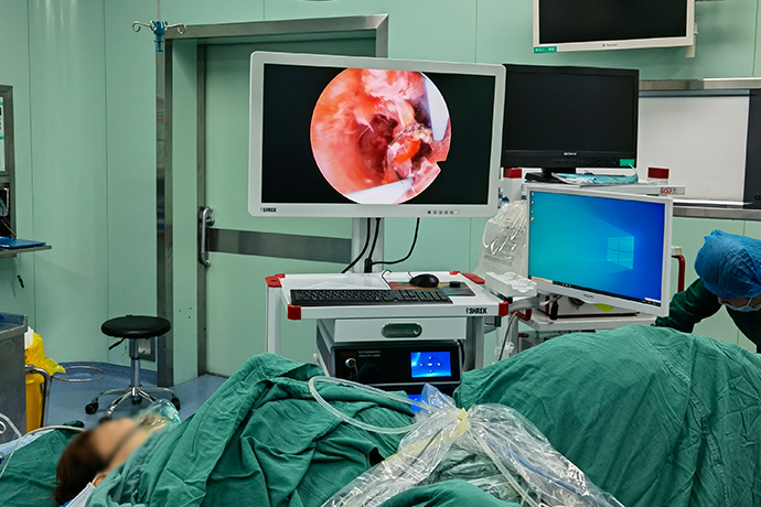
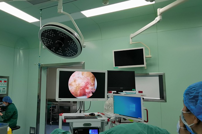
The endoscope, resector and electrodes are assembled and connected to the LED light source, hysteroscope equipment, high-frequency electrosurgical unit and suction irrigation tube. Care must be taken to remove all air bubbles from the pipeline. Then introduce the resectoscope under video guidance, and identify and analyze the number, size, type and precise location of uterine fibroids. Perform hysterectomy using the cutting mode of the electrosurgical device.
• Hysteromyomectomy
• Pedicled fibroids
For obstructive leiomyomas smaller than 20mm (grade 0), the base of the leiomyoma is removed at the level of the healthy endometrial surface under visual guidance. Then a ring (no current) or blunt dissociator must be used to extract the leiomyoma.
For grade 0 leiomyomas larger than 20mm, the gynecologist will gradually remove the leiomyomas under visual guidance, from the free edge to the level of the healthy endometrial surface. During this step, a loop (no current) or a blunt stripper should be used to periodically remove the cutting chips after resection to maintain proper endoscope visibility.
• Grade 1 and 2 leiomyomas
For grade 1 and grade 2 leiomyomas, the gynecologist begins by removing the intracavitary part. After removing the excised shavings with a ring or blunt curette, the boundary between the intramural part of the leiomyoma and the healthy myometrium must be determined (this is easier to discharge, the color is more pink, and not too firm) Therefore, the end point of resection can be selective and does not pursue more than leiomyoma. Several methods can promote the protrusion of the intracavitary part of the leiomyoma in the cavity: massage the leiomyoma with a ring, alternately open and inject 10 IU of oxytocin hydromassage (slow IV) at the same time with a pump. Then complete the resection, always keeping the edge in view.
• Difficult situation
In some more difficult cases (leiomyoma> 40mm, poor visibility, technical problems, operation time> 45 minutes or shortage> 500mL), the gynecologist must leave the base of the leiomyoma and schedule a second operation.
When removing leiomyomas in the uterine cornea, special precautions must be taken because there is a risk of corneal perforation (uterine wall thickness = 3-5mm) and the risk of damaging the uterine tube (must be kept under visual control) women of childbearing age.
In the case of local bleeding, the ablation ring can be used in "clotting mode" to stop bleeding, as long as it is done selectively, because the spread of coagulation may be harmful to the endometrium.
In menopausal patients with multiple endometrium, it is useful to perform hysteroscopic resection of the endometrium (endometrial resection).
Postoperative management
Hysteroscopic myomectomy is performed in an outpatient clinic. No intravenous analgesics are specified.
Only when infertility is an indication for leiomyoma resection, a follow-up diagnostic hysteroscopy is required 2 months after the operation. The purpose of this control is to check postoperative adhesions (10% of cases). It is usually easy to remove these recent small adhesions during the diagnosis of the hysteroscope tip.
If the gynecologist suspects the continuity of the intraluminal fibroma tissue after the removal of the large fibroids to determine whether to indicate a new surgical hysteroscopic surgery, follow-up diagnostic hysteroscopy is also performed.
• Histology
Leiomyomas (also called fibroids) are tumors in which blood vessels made up of smooth muscle fibers surrounded by collagen fibers do not coincide. They are firm and can be calcified. These benign tumors must be distinguished from leiomyosarcoma.
1. The round ligament
2. Fallopian tube
3. The fundus of the uterus
4. Proper ovarian ligament
5. Uterine cavity
6. Endometrium
7. Myometrium
8. Broad ligament mesentery
9. Uterine artery
10. The ureter
11. Cervical canal
12. Uterosacral ligament
13. The outer mouth of the uterus
14. Vagina
• Leiomyoma
Depending on the location of the leiomyoma relative to the largest transverse diameter of the myometrium, three types of leiomyoma can be distinguished:
1. Uterine leiomyoma, located on the surface of the uterus on the peritoneal side;
2. Intermural lymphoma, embedded in the muscle wall of the uterus;
3. Submucosal fibroids, protruding into the uterine cavity;
4. Only submucosal fibroids can be treated by surgical hysteroscopy.
• Submucosal fibroids
According to the degree of intramural development, three levels of submucosal fibroids can be identified:
1. Grade 0: Development is limited to the uterine cavity (with a pedicle or implanted base);
2. Grade 1: Partial intramural development (internal cavity composition> 50%);
Level 3 and 2: Mainly intramural development (lumen<50%).
Indications
-Symptomatic leiomyoma: uterine bleeding, postmenopausal onset, dysmenorrhea, infertility, history of early miscarriage;
-Submucosal positioning (grade 0 and 1; grade 2, if the thickness of the lateral myometrium of the leiomyoma is> 5mm);
-Size <50 mm;
-Single leiomyoma or bifocal positioning, if the size is small.
Contraindications
-Contraindications to anesthesia;
-Genital infections;
-Pregnancy;
-Multiple leiomyomas (indications for open multiple myoma resection).
Preoperative period
• Ultrasonography
Pelvic ultrasound through the abdomen and intravaginal route depicts the size and grade (S) of submucosal fibroids. For grade 2 submucosal fibroids, ultrasound is used to examine the distance between the outer edge of the leiomyoma and the serous membrane of the uterus to assess the risk of intraoperative uterine perforation. Ultrasound is also used to search for related subcutaneous or intramural fibroids.
Saline versus hysteroscopy can also be performed, which involves injecting saline into the uterine cavity during successive scans.
• Diagnostic hysteroscopy
If the ultrasound results are not accurate enough, diagnostic hysteroscopy can confirm the submucosal nature of the fibroids (S). Specify the number, size, type, and location of tumors.
Inform the patient
When planning a hysteroscopic leiomyoma resection, the patient must be informed of the following:
-Risk of complications: uterine perforation, endometritis and metabolic diseases caused by glycine absorption;
-Complete removal may be required during the second operation (40-50mm leiomyoma and grade 2 leiomyoma);
-The possibility of failure of the operation requires a secondary hysterectomy.
Preoperative medication
Preoperative GnRH agonist therapy (gonadotropin releasing hormone) can be prescribed for 2 to 3 months. The goal of this treatment is to reduce the volume of leiomyomas by 30% to 40% before surgery (Friedman et al., 1987; Lawrence et al., 1991). In this case, the procedure should be scheduled 6 to 10 weeks after the first simulated injection. This is the treatment of severe anemia cases specifically marked as GnRH analogs that can treat anemia by suppressing any menorrhagia. In postmenopausal women with cervical stenosis, local or systemic estrogen therapy can be used for the procedure 2 to 3 weeks before surgery to promote cervical dilation. Dilation of the cervix, especially in prenatal patients, can also be achieved by intravaginal administration of misoprostol 2 hours before surgery.
Operating room
• Patients
-General anesthesia or local anesthesia (epidural or spinal anesthesia);
-Stone cutting position;
-Disinfection of the perineum and cervix and vagina;
-Preventive antibiotics when inducing anesthesia to prevent endometritis;
-Urinary catheter (optional).
• Team
1. The surgeon sits in the middle of the patient's leg.
2. The assistant stands on the right side of the surgeon.
3. The anesthesiologist is above the patient's head.
• Equipment
Equipment placed to the left of the surgeon:
-Endoscope and monitor;
-A device that controls the pressure and flow of the expansion medium: a constant uterine expansion must be maintained. The pressure is continuously controlled by suction and irrigation pumps;
-Standard or specially adapted piping for each type of pump;
-Expansion medium: Glycerin solution (1.5% glycine solution packaged in a 3 liter plastic bag) is the most commonly used medium for monopolar cautery. Use bipolar cauterization, use salt water.
-Light source: The same type of xenon light source is used for diagnostic hysteroscopy, surgical hysteroscopy and laparoscopy.
-High frequency electrosurgical unit;
1) Unipolar electrosurgery: use high frequency current (> 300 000 Hz). The division of the tissue is accomplished with unmodulated current that produces a rapid temperature rise.
2) Bipolar electrosurgery: saline is used as a dilation medium to reduce the risk of metabolic complications. The operating channel is narrow, which simplifies the expansion. There are "spray" and "dry" modes. The maximum power used by the generator is 200W.
Instrument
General instruments:
1. Hegar’s dilator (No. 1 to No. 10, diameter increased from 0.5 to 1 mm);
2. Speculum with detachable valve;
3. Resection electrode (4 mm), ending with a 90°cutting circuit (7 to 9 mm), used for monopolar hysteroscopy, or 90°24 French cutting ring or 5 French tip bipolar hysteroscope;
4. Rigid endoscopes have a diameter between 2.7 and 4 mm; the sight direction usually used in hysteroscopy is 12°.
5. Resection endoscope: 7 to 9 mm, with two channels, one internal (flushing) and one external (suction) for monopolar hysteroscopy, or from 5 to 9 mm dual channel and dual current operating channel bipolar hysteroscopy. In all cases, it has an operating handle: passive (electrode input) or active (electrode output);
6. Hysteroscopy;
7. Irrigation and suction channels;
8. Two Pozzi grippers;
9. Uterine measuring device.
System adjustment
Unipolar system
The described ablation technique uses unipolar current. The suction pump must be preset to maintain intrauterine pressure ≤100mm Hg, 250 mL/s flow rate and 0.2 bar suction pressure.
The procedure must not last more than 45 minutes. The total volume of the glycerin solution used must be limited to 6L. The inflow and outflow of the expansion liquid must be accurately monitored. If there is a difference between the flushing flow rate and the suction flow rate, the procedure must be stopped immediately (a 500mL difference can be allowed). If the difference is too large, or if the procedure lasts too long, a chemical test must be performed immediately after the procedure for checking for hyponatremia.
Bipolar system
Bipolar electrospray is a relatively new system. It is non-toxic and the effect seems to be equivalent to that of commonly used unipolar devices. The diameter of the operating channel is the same as the new 24 French cutting cycle and a smaller bipolar system when using 5 French bipolar tips to avoid the need for expansion. The swelling medium used is saline, which reduces the risk of metabolic complications and allows the procedure to last longer. With the French 24-electrode resection, the operation steps and adaptations for the type of uterine fibroids are the same. The hysterectomy adopts a unipolar system. 5 The French bipolar system can be used for fibroids less than 20 mm in diameter. The technique of this system includes contacting and spraying the base of the uterine fibroids, if it is pedicled, or if the entire fibroids are unstemmed. In theory, bipolar systems are safe because they can interact with salt water. In contrast to the unipolar system, which penetrates into the tissue, it can be partially concealed at certain points, and the bipolar system is often visible.
Surgical steps
• Cervical dilation
A two-handed examination is performed to evaluate the position of the uterus before dilation. This reduces the risk of perforation. Insert a speculum with a detachable valve, and grasp the cervix with 2 Pozzi or Museux-Palmer graspers placed at the 3 o'clock and 9 o'clock positions to bring the uterus into the middle position. If no diagnostic hysteroscopy is performed during the preoperative evaluation, the process usually starts with the diagnostic hysteroscopy. Then use the Hegar dilator to dilate the cervix, using gradually increasing dilators, until a size 10 dilator can be inserted.
• Insert the resectoscope
The endoscope, resector and electrodes are assembled and connected to the LED light source, hysteroscope equipment, high-frequency electrosurgical unit and suction irrigation tube. Care must be taken to remove all air bubbles from the pipeline. Then introduce the resectoscope under video guidance, and identify and analyze the number, size, type and precise location of uterine fibroids. Perform hysterectomy using the cutting mode of the electrosurgical device.
• Hysteromyomectomy
• Pedicled fibroids
For obstructive leiomyomas smaller than 20mm (grade 0), the base of the leiomyoma is removed at the level of the healthy endometrial surface under visual guidance. Then a ring (no current) or blunt dissociator must be used to extract the leiomyoma.
For grade 0 leiomyomas larger than 20mm, the gynecologist will gradually remove the leiomyomas under visual guidance, from the free edge to the level of the healthy endometrial surface. During this step, a loop (no current) or a blunt stripper should be used to periodically remove the cutting chips after resection to maintain proper endoscope visibility.
• Grade 1 and 2 leiomyomas
For grade 1 and grade 2 leiomyomas, the gynecologist begins by removing the intracavitary part. After removing the excised shavings with a ring or blunt curette, the boundary between the intramural part of the leiomyoma and the healthy myometrium must be determined (this is easier to discharge, the color is more pink, and not too firm) Therefore, the end point of resection can be selective and does not pursue more than leiomyoma. Several methods can promote the protrusion of the intracavitary part of the leiomyoma in the cavity: massage the leiomyoma with a ring, alternately open and inject 10 IU of oxytocin hydromassage (slow IV) at the same time with a pump. Then complete the resection, always keeping the edge in view.
• Difficult situation
In some more difficult cases (leiomyoma> 40mm, poor visibility, technical problems, operation time> 45 minutes or shortage> 500mL), the gynecologist must leave the base of the leiomyoma and schedule a second operation.
When removing leiomyomas in the uterine cornea, special precautions must be taken because there is a risk of corneal perforation (uterine wall thickness = 3-5mm) and the risk of damaging the uterine tube (must be kept under visual control) women of childbearing age.
In the case of local bleeding, the ablation ring can be used in "clotting mode" to stop bleeding, as long as it is done selectively, because the spread of coagulation may be harmful to the endometrium.
In menopausal patients with multiple endometrium, it is useful to perform hysteroscopic resection of the endometrium (endometrial resection).
Postoperative management
Hysteroscopic myomectomy is performed in an outpatient clinic. No intravenous analgesics are specified.
Only when infertility is an indication for leiomyoma resection, a follow-up diagnostic hysteroscopy is required 2 months after the operation. The purpose of this control is to check postoperative adhesions (10% of cases). It is usually easy to remove these recent small adhesions during the diagnosis of the hysteroscope tip.
If the gynecologist suspects the continuity of the intraluminal fibroma tissue after the removal of the large fibroids to determine whether to indicate a new surgical hysteroscopic surgery, follow-up diagnostic hysteroscopy is also performed.
- Recommended news
- 【General Surgery Laparoscopy】Cholecystectomy
- Surgery Steps of Hysteroscopy for Intrauterine Adhesion
- [Percutaneous Endoscopic Lumbar Discectomy] Percutaneous endoscopic lumbar discectomy for lumbar disc herniation
- [Urology Cystoscopy Section] Cystoscopic Laser Lithotripsy
- [Laparoscopic Urology] Laparoscopic Radical Right Nephrectomy