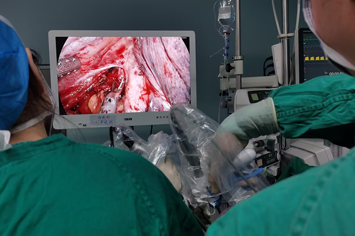
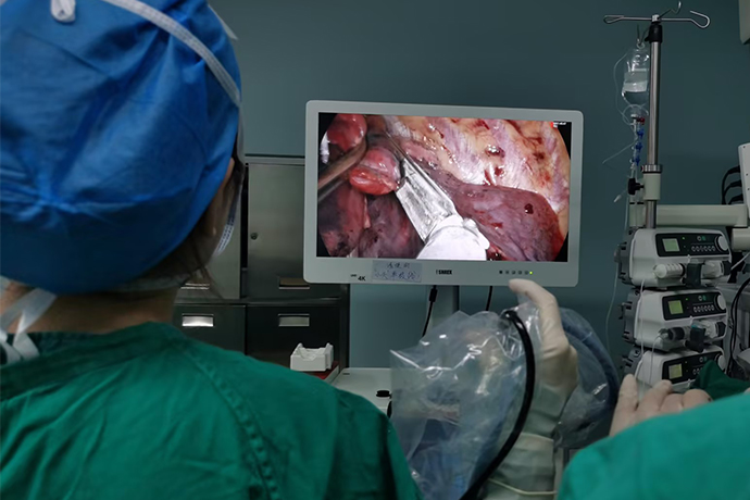
[Thoracoscopic surgery of thoracic surgery] 4K ultra-high definition pulmonary metastatic nodule resection
Release time: 26 Dec 2023 Author:Shrek
As we all know, the human lungs are extremely rich in blood, and the blood throughout the body is oxygenated here and then participates in circulation. With such a fertile "land", many cancer cells will naturally not miss it, and unknowingly expand their territory to the lungs, such as breast cancer, liver cancer, colorectal cancer, renal cell cancer, etc. Intrapulmonary metastasis of these extrapulmonary malignant tumors may be a sign of spread of the disease. Some patients may have pulmonary metastases without evidence of the primary tumor. I would like to know what are the symptoms of pulmonary metastases and what to do. Treatment, can surgery be performed? Let’s watch it together!

Are there any "clues" to follow for lung metastases?
In the early stages of lung metastasis, lung metastasis tumors have few symptoms, and metastases are mostly discovered through imaging examinations, such as CT or PET/CT scans.
Some patients develop symptoms that are often similar to those of primary lung cancer, especially patients with hilar involvement who are prone to persistent cough. Common symptoms include coughing up blood, hemoptysis or blood-streaked sputum, persistent chest, shoulder and back pain, as well as shortness of breath, hypoxemia, pleural effusion, etc. Other symptoms: if the tumor compresses the superior vena cava, it can lead to superior vena cava syndrome. The patient will present with edema of the head, face and upper limbs, dizziness, and dilation of superficial veins in the neck and upper chest (veins that are not originally exposed are significantly expanded and exposed)
The presence of metastatic cancer means that the primary cancer may have spread throughout the body, so general symptoms of cancer are also common, such as fatigue, unexplained weight loss, or loss of appetite.
How to treat lung metastases?
How to treat lung metastases usually depends on the source of the primary tumor. These treatments can include endocrine therapy, targeted therapy, chemotherapy, immunotherapy or multidisciplinary treatment.
For patients with advanced cancer, chemotherapy is usually used as palliative treatment in order to reduce symptoms and improve quality of life. But sometimes surgical therapy is chosen. Large-scale clinical trials have proven that resection of many pulmonary metastases of different malignant origins can prolong survival and even have a curative effect. Therefore, it can be said that active resection of isolated pulmonary metastases is A proven method.
Although most of the current clinical data are retrospective analyzes and lack the results of randomized trials, the results show that active surgical treatment is helpful in prolonging the survival of patients.
Six indications for surgical resection of pulmonary metastases
There are currently few published guidelines on the surgical indications for metastasectomy. The NCCN guidelines and the 2018 American Society of Thoracic Surgeons consensus document are available.
The criteria for resection can be summarized as follows:
1. According to preoperative imaging results, pulmonary metastases can be completely resected
2. The patient’s cardiopulmonary function can tolerate the surgery
3. Metastasectomy is technically feasible
4. The primary tumor is controllable
5. There is no extrapulmonary metastasis and extrapulmonary metastasis is controllable
6. Other special circumstances: For metastatic tumors accompanied by hilar and mediastinal lymph node metastasis, it is often a sign of systemic metastasis. The surgical effect is not ideal and surgical treatment is not recommended. In addition, the possibility of primary tumor must be excluded, and further histopathological diagnosis is required.
Surgery for metastatic lung cancer
The specific surgical method selected for metastatic lung cancer should be based on the size, location, and extent of invasion of the metastatic tumor. The main surgical method is the same as that for primary lung cancer, which is partial lung resection, including wedge resection and segmental resection. Wedge resection is a classic local resection method with less trauma and faster recovery.
With the continuous development and maturity of 4K ultra-high-definition thoracoscopy technology, minimally invasive surgery for early lung cancer has made great progress in the past 20 years. Currently, video-assisted thoracoscopic surgery has become the main surgical treatment for lung cancer. The results of clinical multi-center studies at home and abroad show that 4K ultra-high-definition thoracoscopic surgery can achieve the same effect as traditional thoracotomy in treating lung cancer.
4K ultra-high-definition thoracoscopy surgery opens 2 to 3 holes in the chest wall without using retractors or ribs. The chest cavity is observed through the 4K ultra-high-definition medical monitor screen, and blood vessels and bronchi are ligated separately, and hilar and mediastinal lymph nodes are removed or sampled. . It has the advantages of small incision, light pain, beautiful appearance, and quick postoperative recovery.
1. Differences from traditional open surgery
(1) Different sizes of incisions
The disadvantages of traditional surgery are that the surgical incision is too long, the latissimus dorsi muscle and serratus anterior muscle need to be cut off, the trauma is large, the bleeding is large, the chest opening and closing time is long, the postoperative recovery is slow, the hospitalization time is long, especially the postoperative wound pain is severe, and it is easy to Lead to the occurrence of complications such as cardiovascular and respiratory systems. Minimally invasive surgery only requires making 2-3 holes in the chest wall, and the lesions can be removed without using retractors or ribs. The trauma during the surgery is light, the bleeding is small, the impact on the cardiopulmonary function is small, and the patient recovers quickly.
(2) The perspective of the surgeon is different
Using a tiny 4K ultra-high-definition medical camera to project the situation inside the chest onto a large medical display screen is equivalent to putting the doctor's eyes into the patient's chest to perform surgery. The surgical field of view can be enlarged as needed to display subtle structures, which is clearer and more flexible than direct vision with the naked eye. Therefore, the exposure of the surgical field of view, the appearance of the microstructure of the disease, the judgment of the scope of surgical resection, and the safety are better than open surgery.
(3) The surgical instruments are different. 4K ultra-high-definition minimally invasive thoracoscopic surgery requires the help of endoscopic linear cutting staplers, electrosurgical scalpels, ultrasonic scalpels and other instruments.
Because the surgical incision is small, special endoscopic cutting and suturing devices and other endoscopic instruments (scissors, separation forceps, etc.) need to be used. Multiport minimally invasive surgery can use instruments through the secondary operating holes to assist in exposure and separation; single-port minimally invasive surgery requires the use of extended curved double-joint instruments that can be entered through the incision at the same time to cross-operate the instruments without "fighting". It places higher demands and challenges on the surgeon's minimally invasive skills, touch, patience and meticulousness.
(4) Suitable patient groups are different
1. Minimally invasive methods are more suitable for early stage treatment, and the tumors are small, especially ground glass nodules and pulmonary nodules are absolute indications.
At present, the ideal screening conditions for minimally invasive surgery patients are early stage, peripheral nodules, no obvious adhesions in the chest, no calcified adhesions of lymph nodes in the hilus, and clear anatomy.
2. Open for mid-term central type, large mass
Generally speaking, it is best not to undergo minimally invasive surgery for tumors larger than 7 centimeters. Even if minimally invasive surgery is performed, the specimen cannot be taken out, and a chest opening is required to collect the specimen. At present, some people think that the specimen should be broken into pieces and then taken out. There is a bag for taking the specimen and the pieces should be broken in this bag. However, a tumor that is too large will affect the doctor's operation. Second, if the adhesions in the chest are particularly severe or the anesthesia is difficult, thoracoscopy cannot be performed. In addition, there is a close relationship between tumors and blood vessels. It is estimated that bleeding may occur during thoracoscopic surgery. In this case, it is best to perform thoracotomy for safety reasons.
2. Types of minimally invasive surgery
Minimally invasive thoracoscopic surgery: 4 holes, 3 holes, 2 holes, single hole.
Both single-port minimally invasive surgery and multi-port minimally invasive surgery are optional surgical approaches in minimally invasive lung surgery. Surgeons should choose different surgical approaches based on their own habits, patient conditions or other special circumstances, and should not stick to one. All choices should be based on the patient's safety and the principle of complete tumor resection.
3. Advantages
(1)Small incision, light pain, beautiful appearance.
Only 1-3 small holes with a length of 1.5 cm are made on the chest wall. Compared with the traditional thoracotomy incision of about 30 cm, the patient's pain and surgical risk can be significantly reduced. The postoperative recovery is faster and the wound is more beautiful.
(2)4K ultra-high-definition thoracoscopy surgery has good magnification effect. 4K ultra-high-definition thoracoscopy surgery has clear vision.
The minimally invasive thoracic surgeon performed local anesthesia on the patient under CT guidance. The relatively superficial right upper lung nodule was punctured into the lung surface with a puncture needle and then injected with methylene blue staining for marking. The left upper lung nodule was deeper and larger. Use a lung puncture positioning needle and hook the tip of the positioning needle into the lung tissue around the nodule.
4K ultra-high-definition single-port thoracoscopic surgery is performed under general anesthesia, and precise resection is performed based on the position of the nodule under the microscope. This type of surgery removes preoperative preparation, positioning, anesthesia, thoracotomy, chest closure and other steps. The removal process only takes a few minutes, the wound is 2-3cm, and the patient can be discharged from the hospital in 3 days.

- Recommended news
- 【General Surgery Laparoscopy】Cholecystectomy
- Surgery Steps of Hysteroscopy for Intrauterine Adhesion
- [Otolaryngology Nasal Endoscopy] How to Treat Recurrent Rhinitis
- [ENT Surgery: Nasal Endoscopy] Endoscopic Treatment of Nasal Polyps
- [Otolaryngology Nasal Endoscopy] Methods and Precautions for Nasal Bleeding Control under Nasal Endoscopy