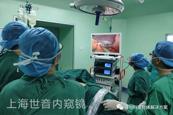
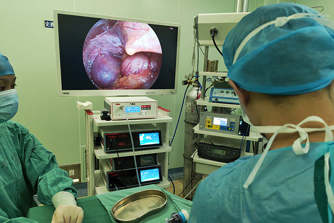
[Laparoscopic Hepatobiliary Surgery] Splenectomy
Release time: 04 Jan 2023 Author:Shrek
Splenectomy is widely used in splenic trauma, splenic abscess, splenic tumor, splenic cyst, gastric cancer, intrahepatic portal hypertension combined with hypersplenism and other diseases. With the continuous development of endoscopic surgical techniques, laparoscopic splenectomy has been successfully applied. Due to its advantages of minimal trauma, less pain, faster recovery and shorter hospital stay, it has developed rapidly. Now laparoscopic splenectomy can be applied to the vast majority of diseases that require surgical removal of the spleen, including hematological diseases, benign and malignant spleen tumors, splenic cysts, free spleen, and AIDS splenectomy.

Preoperative preparation
(1) Liquid diet 1 day before operation, intestinal preparation, fasting in the morning of operation, placement of nasogastric tube, skin preparation, etc.
(2) Prophylactic antibiotics should be applied 1 to 2 days before operation, and 1 to 2 weeks before operation for immunocompromised patients.
(3) Liver function should be improved, bleeding tendency and anemia should be corrected before operation.
(4) Equipment preparation.
The spleen is a solid organ rich in blood supply, soft and brittle, dark red in color. The spleen is located deep in the left hypochondrium, on the left side of the stomach, below the diaphragm, approximately parallel to the tenth rib. The entire spleen is covered by the 9th, 10th, and 11th ribs.
The splenic artery comes from the celiac artery, and before entering the splenic hilum, it divides into upper and lower branches or three upper, middle, and lower branches, and then divides into secondary or tertiary branches to enter the splenic hilum.
There are about 2 to 4 splenic veins, coiled and accompanied by the splenic artery, and flow into the main trunk outside the splenic hilum.
Gastrosplenic ligament: Between the medial front of the spleen and the greater curvature of the stomach, there are short gastric arteries, veins, and left gastroepiploic arteries and veins; the upper part of this ligament is often shorter, making the upper pole of the spleen very close to the greater curvature of the gastric fundus , When cutting this ligament in surgery, if you are a little careless, it is easy to damage the stomach wall.
Gastrosplenic ligament: It is the double-layered peritoneum from the hilum of the spleen to the front of the left kidney, containing the splenic artery and its branches, the splenic vein and its subordinate branches, and splenic lymph nodes. Sometimes the tail of the pancreas protrudes, so do not injure the tail of the pancreas when handling the blood vessels of the splenic pedicle during splenectomy.
Spleenorenal ligament: It is the double-layered peritoneum from the hilum of the spleen to the front of the left kidney, containing the splenic artery and its branches, the splenic vein and its branches, and splenic lymph nodes. Sometimes the tail of the pancreas protrudes, so do not injure the tail of the pancreas when handling the blood vessels of the splenic pedicle during splenectomy.
Spleenocolic ligament: short, connecting the inferior spleen and the splenic flexure of the colon. Be careful not to injure the colon during splenectomy.
Diaphragmatic splenic ligament: It connects the upper pole of the spleen and the peritoneum between the spleen, is relatively narrow, and is only seen in a few peritoneal folds formed at the corner of the upper pole of the spleen to the visceral surface. This ligament is more prominent in splenomegaly.
Indications for splenectomy
1. Significant enlarged spleen (giant spleen), causing obvious symptoms of abdominal compression.
2. Combined with severe anemia, especially hemolytic anemia; thrombocytopenia (PLT less than 10*10E9/L) or spontaneous bleeding tendency; white blood cell or agranulocytosis (white blood cell less than 1.0*10E9/L).
3. Comprehensive treatment measures such as liver cirrhosis complicated with esophageal variceal bleeding, drug therapy, repeated endoscopic therapy, and interventional therapy are ineffective or unavailable.
4. The general condition and liver function permit, especially the child A or B+ liver function.
Key surgical steps
The improved lithotomy position was taken, the pneumoperitoneum was established, the operation hole was established, the spleen was dissociated, the spleen pedicle was treated, the spleen was taken out, fixed with sutures, the pneumoperitoneum was closed, and the puncture hole was sutured.
1. Posture
Position the patient securely on the bean bag with the left side tilted upward 60° in supine inversion with the head down and the left arm positioned for the left thoracotomy. This allows gravity to retract the abdominal organs and maximize working space. The surgeon stands on the right side of the patient facing the left monitor, and the camera assistant on the same side sits on a stool to his left to avoid conflict with the surgeon's elbow.
2. Selection of puncture hole location
3. Abdominal exploration
When performing splenectomy for chronic diseases, it is necessary to investigate the cause of the preoperative diagnosis, including the size of the spleen, whether there is adhesion, whether there is an accessory spleen, the relationship with the surrounding organs, and the condition of the splenic artery and vein. In cases of cirrhotic portal hypertension, attention should be paid to the size of the liver, the degree of cirrhosis, the presence or absence of new organisms, the amount of collateral circulation in the portal vein system, and the presence or absence of embolism, and the portal vein pressure should be measured. For cases of congenital hemolytic anemia, the gallbladder and bile duct should be detected to determine whether there are stones.
4. Surgical procedure
The approach of splenectomy is generally divided into anterior approach (splenogastric ligament→splenocolic ligament→splenorenal ligament→splenophrenic ligament→splenic pedicle) and posterolateral approach (splenorenal ligament→splenocolic ligament→splenogastric ligament→splenic diaphragm). ligament → spleen pedicle). Here, the previous approach is taken as an example to explain the operation process in detail.
Severe the spleen-gastric ligament, ligate the short gastric vein and the exposure of the pancreatic tail.
Lower exposure of the spleen and separation of lower pole vessels.
Treatment of the major vessels of the spleen.
Put the spleen into the retrieval bag and take out the spleen.
Common Complications
Intra-abdominal hemorrhage
Usually occurs within 24-48 hours after surgery. The most common cause is severe hemorrhage from the diaphragm surface, detachment of the splenic pedicle ligation thread, or hemorrhage from vessels that were not ligated during the operation.
Preventive measures: In the operation, repeatedly check the diaphragm surface, the ligated end of the spleen and stomach ligament, the lateral abdominal wall, the retroperitoneum, the spleen pedicle and the tail of the pancreas to see if there are bleeding points, and strictly stop the bleeding.
Subphrenic abscess
Within 1 to 2 weeks after splenectomy, patients often have hypothermia, generally not exceeding 38.5°C. However, if the high fever persists after the operation, or if the body temperature drops and then rises after 1 week after the operation, it cannot be simply regarded as splenectomy fever. More than subdiaphragmatic hemorrhage or infection.
Preventive measures: Strict hemostasis, avoid contusion of the tail of the pancreas when dealing with the splenic pedicle, routinely place effective drainage under the diaphragm after operation, and timely drain the accumulated blood in the splenic fossa, all of which are effective measures to prevent subdiaphragmatic abscess.
Thrombo-embolic complications
Once it occurs in some parts of the blood vessels, such as retinal artery, mesenteric vein, portal vein trunk, etc., it will cause serious consequences. The occurrence of this complication is related to the rapid increase in platelet count after splenectomy.
Preventive measures: most of them advocate the application of anticoagulants such as heparin for preventive treatment when the platelet count exceeds 1000~2000*10⁹/L after splenectomy. If thrombo-embolic complications occur, anticoagulant therapy should be used, and treatment and rest, aspirin, dipyridamole and other drugs can also be added.
Postoperative care
1. In addition to the post-treatment of the general abdominal surgery, those with damage to liver function should also actively adopt liver protection therapy.
2. If there is a drainage tube, it can be connected to a bedside disinfection bottle, and the drainage tube will be removed 3 days after the operation.
3. Generally, you can leave the hospital 3 days after the operation.
Precautions
If severe splenic adhesions are found during the operation, giant splenectomy is difficult, or massive bleeding occurs during the operation and laparoscopy cannot quickly stop the bleeding, it should be transferred to laparotomy or changed to one-handed assisted laparoscopic splenectomy.

- Recommended news
- 【General Surgery Laparoscopy】Cholecystectomy
- Surgery Steps of Hysteroscopy for Intrauterine Adhesion
- [Otolaryngology Otoscopic Section] Excision of Cholesteatoma in the External Auditory Canal
- [Otolaryngology Nasal Endoscopy] How to Treat Recurrent Rhinitis
- [ENT Surgery: Nasal Endoscopy] Endoscopic Treatment of Nasal Polyps