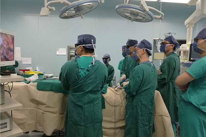
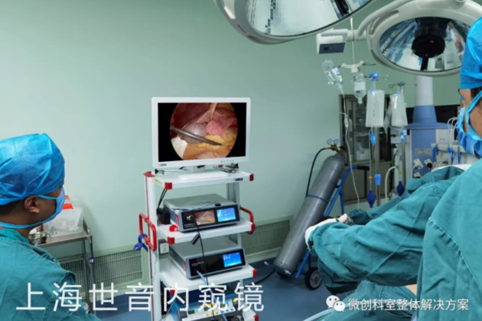
[Laparoscopic Hepatobiliary Surgery] Left lateral hepatic lobectomy
Release time: 13 Dec 2022 Author:Shrek
Laparoscopic left lateral hepatectomy
Laparoscopic surgery has the advantages of less trauma, faster recovery, fewer complications, and shorter hospital stay, which is significantly better than open surgery, and has become a trend in the world today. Laparoscopic left lateral segment liver resection is the basic operation of laparoscopic liver lobectomy. It is the earliest laparoscopic liver resection with mature technology. It is easy to learn and master, and has been widely carried out in major hospitals. . The hepatic blood flow and bile drainage of the left outer lobe of the liver are in the I/M segment hepatic pedicle, and the hepatic blood flow is in the left hepatic vein. Therefore, as long as the I/M segment hepatic pedicle and the left hepatic vein are properly handled, the abdominal cavity can be successfully completed. Microscopic left lateral hepatectomy. According to different surgical instruments and surgical methods, surgeons innovatively explored the surgical method of laparoscopic left lateral hepatectomy in clinical practice. Anatomic hepatectomy can be performed, and irregular hepatectomy can be adopted. Dr. Zhang Jihong's team flexibly applied the two methods to achieve the effect of bloodless liver resection and ensure the safety of the operation to the greatest extent. Combining with literature review, academic technology exchange and own practical experience, the relevant knowledge of laparoscopic left lateral hepatectomy is briefly described as follows. Be sure to refer to.

Potential advantages of laparoscopic liver resection
Operation
Reduce postoperative pain
Early postoperative mobilization
Enhance lung function during surgery
Reduce wound complications
Relief of perioperative immunosuppression
The unique anatomical structure of the left lateral lobe of the liver makes laparoscopic left lateral hepatectomy an earlier and more widely used laparoscopic hepatectomy. The operation time of this operation is comparable to that of laparotomy, but due to the small surgical incision and less trauma, it is significantly better than laparotomy in terms of short-term prognosis. Today I share a case about laparoscopic left lateral hepatectomy.
Case information
A hyperechoic hypovascular mass of 5.1 cm in the left hepatic lobe was found incidentally during CT urography for renal stones.
History of long-term contraceptive use.
Laparoscopic appendectomy and tubal cyst resection were performed 3 years ago.
A diagnosis of HNF1α-inactivated hepatic adenoma was made.
Occurs only in women, especially women taking oral contraceptives.
50% of patients had multiple tumors.
The risk of intramural hemorrhage and rupture increases when the tumor size is greater than 5 cm.
The risk of malignant transformation is low.
The lesion usually resolved after discontinuation of oral contraceptives, but the patient remained unchanged in size and a laparoscopic left lateral hepatectomy was planned.
Surgical procedure
1. Preliminary examination of the abdomen revealed 10 well-defined masses in the S2 and S3 segments of the liver.
2. Use the ultrasonic scalpel to cut the round ligament, falciform ligament, left triangular ligament and left coronary ligament sequentially.
If there is a large blood vessel in the left triangular ligament, clip it and then cut it off. The assistant lifts the left outer lobe of the liver, and the ultrasonic knife dissects the lesser omentum until near the root of the venous ligament (some patients may not need to deal with the lesser omentum). Sufficient dissociation is crucial for subsequent operations. During the operation, there is no need to intentionally expose the left hepatic vein or inferior vena cava outside the liver.
3. From the falciform ligament into the capsule of Glisson.
Start along the round ligament of the liver and the left edge of the falciform ligament, from the foot side to the head side, from shallow to deep, and gradually proceed.
4. Ultrasonic scalpel and endovascular stapler were used to separate the liver parenchyma.
The upper and lower liver tissues of the S2 and S3 Glisson sheaths were cut to initially expose the S2 and S3 Glisson branches. When the liver parenchyma was severed, the larger ducts were closed with surgical clips, titanium clips or absorbable clips before cutting off.
After dissecting the Glisson pedicle of the S2 and S3 segments, continue to dissect the liver parenchyma toward the root of the left hepatic vein, cut off the liver tissue above and below the left hepatic vein, expose the root of the left hepatic vein, and dissect the left hepatic vein with a linear cutting occluder. Be careful not to injure the diaphragm.
5. The cut surface of the liver was cauterized with an argon knife until a satisfactory hemostasis effect was obtained.
6. Dilute hydrogen peroxide to check for bile leakage and bleeding.
7. Take out the specimen from the Pfannenstiel's incision on the suprapubic abdomen, cut open and suture the skin.
Hemostasis and prevention of bile leakage are the main purposes of section management after hepatectomy. For bleeding from liver wounds, bipolar coagulation or argon knife can be used to stop bleeding; for active bleeding, non-invasive sutures should be used to stop bleeding. After the author washed the wound with normal saline, no obvious bleeding or oozing was seen, and no hemostatic gauze was placed. A single-lumen drainage tube was placed under the wound surface at the lateral border hole of the right rectus abdominis muscle. Considering the postoperative aesthetics, the author used 3-0 vascular sutures for intradermal sutures. Compared with absorbable sutures, vascular sutures have a lower risk of foreign body reactions. Vascular sutures can be removed after 1 week.
Placement of single-lumen drainage tube and hepatic hemangioma tissue
In order to maintain a clear view of the laparoscopic operation, bleeding from the abdominal cavity or liver wound should be stopped as soon as possible to avoid causing massive bleeding and increasing the risk of conversion to laparotomy. Intraoperative ultrasonography plays an important role in evaluating the resectability of malignant tumors. Intraoperative ultrasonography can not only clarify the size and location of lesions, improve the radical resection of surgery, but also determine the direction of important pipelines, effectively avoid damage, and reduce the risk of surgery. The risk of severe bleeding and the like. For benign tumors, intraoperative ultrasound is not necessary. Following the no-touch principle of tumors, the application of specimen bags can reduce the incidence of tumor implantation and metastasis.
Postoperative results
Very little blood loss.
The operation time was 159 minutes.
Postoperative recovery was smooth.
Discharged on the 2nd postoperative day.
The final pathological diagnosis was hepatocellular adenoma, HNF1 α-inactive type.
Surgical margins were negative and there was no malignancy.

- Recommended news
- 【General Surgery Laparoscopy】Cholecystectomy
- Surgery Steps of Hysteroscopy for Intrauterine Adhesion
- [Otolaryngology Otoscopic Section] Excision of Cholesteatoma in the External Auditory Canal
- [Otolaryngology Nasal Endoscopy] How to Treat Recurrent Rhinitis
- [ENT Surgery: Nasal Endoscopy] Endoscopic Treatment of Nasal Polyps