[Orthopedic UBE Section] Four Years of Evolution of UBE Technology: From Lumbar Fusion to the "Forbidden Zone" of Thoracic and Cervical Spine, and the Unignorable "Hydraulic Pressure Crisis"
Release time: 01 Sep 2026 Author:Shrek
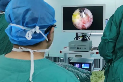
The advancement of minimally invasive spinal surgery has never stopped, from the initial open surgery to microsurgery with portal assistance, and now to ultra-minimally invasive endoscopic techniques. Along this evolutionary path, unilateral biportal endoscopy (UBE), also known as bilateral endoscopic spinal surgery (BESS), has emerged as a disruptive force between 2020 and 2024.
Unlike the single-port endoscope (PELD/FESS) which uses a single channel for simultaneous observation and manipulation, the UBE employs a "floating" dual-channel system: one channel is used for arthroscopic observation, and the other serves as a separate working channel. This separate design allows surgeons to freely use standard open surgical instruments such as drills, scalpels, and forceps under clear water-based visualization.
The past four years have been a period of "maturation" for UBE (Underlying Belt Embolization) technology. It is no longer merely an alternative to lumbar discectomy, but has undergone rigorous clinical validation, venturing into multi-segmental spinal stenosis, lumbar spondylolisthesis fusion, and even high-risk ossification of the ligamentum flavum in the thoracic spine.
However, with the aggressive expansion of indications, a new physiological challenge has emerged—hydrodynamic risks. Continuous saline perfusion not only provides clear visualization but also introduces potential complications such as increased intracranial pressure and seizures.
This article will summarize the latest research hotspots, expanded clinical indications, competition with the "gold standard," and crucial fluid safety issues related to UBE technology, based on core global literature from 2020 to 2024.
I. Global Perspective: From "Niche" to "Mainstream"
Before 2020, UBE was largely considered an emerging experiment. However, bibliometric analysis shows that academic output in this field has exploded since 2019-2020, peaking in 2023. Even during the COVID-19 pandemic, the global demand for shorter hospital stays and day surgery in spine surgery has accelerated the adoption of UBE (Ultra-East Asian Endoscopy) technology.
Establishment of the "East Asian Axis"
Currently, UBE research exhibits a clear East Asian dominance:
• South Korea: As a pioneer and innovation engine for this technology, South Korea contributed approximately 70% of the early literature and boasts numerous global training centers (such as Wooridul Spine Hospital).
• China: Chinese doctors have followed up remarkably quickly, contributing significantly in recent years in terms of case volume and high-quality comparative studies (meta-analysis). Clinical practice in China and South Korea has effectively defined the global standard for this technology.
II. Learning Curve: Easier to Learn Than Single-Port Endoscopy?
The core advantage of UBE lies in the "triangulation" principle. Independent observation and working channels grant surgeons a high degree of operational freedom, very similar to the logic of traditional microsurgery or knee arthroscopy.
1. A Flatter Learning Curve
The consistent conclusion of Cumulative and United States Analysis (CUSUM) studies is that the learning curve for UBE is flatter than that for single-port PELD (Pelt-Off Lamp). • Proficiency Threshold: Surgical time and complication rates typically stabilize after 28-35 surgeries.
• Comparative Advantage: In contrast, single-port PELD, due to its two-dimensional monocular field of view and "lever effect," often requires a longer adaptation period (35-40+ cases) to rebuild hand-eye coordination.
2. Stage-Specific Characteristics
• Early Stage (1-25 cases): Common problems include long surgical time, frequent fluoroscopy sessions, and "collision" (interference) between the two instruments within the spinal canal.
• Mature Stage (>35 cases): Decompression surgery time typically stabilizes at 60-65 minutes.
III. Breakthrough in Indications: UBE Lumbar Interbody Fusion (ULIF) The most significant clinical trend over the past four years has been the widespread application of UBE in lumbar interbody fusion (ULIF). This represents a deep integration of endoscopic techniques and reconstruction concepts.
1. Technological Advantages
Unlike single-port endoscopic fusion (ULIF), which often requires a special expandable fusion device, ULIF allows surgeons to implant a large-diameter conventional fusion device and percutaneous pedicle screws through the working channel. More importantly, under direct endoscopic visualization, surgeons can more thoroughly prepare the endplate and remove the cartilaginous endplate, thus improving the fusion rate.
2. ULIF vs. MIS-TLIF (Minimally Invasive Transforaminal Lumbar Interbody Fusion)
Numerous comparative studies have revealed subtle differences in clinical outcomes between the two:
• Fusion Rate: Both are comparable (2-year fusion rates are around 95%-97%), and UBE does not sacrifice biological stability.
• Blood Loss: ULIF is significantly lower (approximately 50 mL vs. 100 mL). This is due not only to the suction device but also to the hemostatic effect of water pressure compressing the epidural venous plexus.
• Procedure Time: MIS-TLIF is generally faster. ULIF, involving dual-channel establishment and water flow management, often extends the surgical time by 20-40 minutes.
• Postoperative recovery: ULIF patients have shorter hospital stays and less early muscle pain.
Expert commentary: The core value of ULIF lies in its superior soft tissue protection and reduced perioperative blood loss, but surgeons need to weigh the extended anesthesia time against the benefits of minimally invasive surgery.
IV. Challenging Forbidden Zones: High-Difficulty Applications to the Cervical and Thoracic Spine
From 2020 to 2024, UBE successfully broke through the "lumbar barrier," venturing into the cervical and thoracic spine, which carry higher risks to spinal cord function.
1. Cervical Spine: Posterior Foraminal Discectomy (UBE-PCF)
For radiculopathy of cervical spondylosis, traditional posterior surgery (Open PCF) is often hampered by postoperative axial neck pain caused by muscle dissection. UBE-PCF provides a solution that preserves the posterior tension band.
• Clinical outcomes: Improvement in the Neck Disorder Index (NDI) is comparable to that of open surgery.
• Refined Approach Selection: Recent research has compared the "ipsilateral approach" with the "contralateral oblique approach." Results show that the contralateral approach has unique advantages: it allows surgeons to vertically observe the nerve root exit zone without damaging the facet joints on the affected side, making it ideal for severe cases of intervertebral foraminal stenosis.
2. Thoracic Spine: Safety Strategies for Ossification of the Ligamentum Flavum (OLF)
Surgery for ossification of the ligamentum flavum (T-OLF) in the thoracic spine carries extremely high risks, with even minor errors potentially leading to paralysis. The application of UBE in this area has undergone a key technological iteration:
• Early Lessons (Inside-Out): Attempting to remove the ossified mass from the spinal canal from the inside out is extremely dangerous and can easily damage the spinal cord.
• Current Standard (Outside-In/Floating): The "whole-piece floating method" is recommended. This involves first thinning the lamina until the ossified mass is completely separated from the bone, appearing "floating," before lifting it entirely from the dura mater. This technique minimizes the compression of the spinal cord caused by instruments entering the spinal canal.
V. The Ultimate Showdown: UBE vs. PELD vs. Microscopy
In the debate over "one hole" versus "two holes," the literature provides a clear picture:
1. UBE vs. PELD (Single-Port):
• Efficiency: For simple soft disc herniation, PELD remains the most efficient, offering faster surgery.
• Capability: For complex spinal stenosis, bony compression, or fusion surgeries, UBE wins. UBE allows for more flexible bilateral decompression (ULBD) and is not limited to a single perspective.
2. UBE vs. Microsurgery:
• Muscle Protection: This is a quantifiable victory. Postoperative creatine kinase (CK) levels and inflammatory markers (CRP) in UBE patients were significantly lower than in the microsurgery group, demonstrating extremely low damage to the multifidus muscle.
VI. A New Risk to Watch Out For: Hydrodynamics and the "Water Pressure Crisis"
With the increasing number of cases, a type of complication rare in open surgery but unique to UBE has attracted significant attention from the academic community—fluid-related complications.
1. Increased Intracranial Pressure and Seizures
The epidural space of the spine is not an isolated island. High-pressure irrigation compresses the dural sac, forcing cerebrospinal fluid (CSF) to shift cephalad, thus increasing intracranial pressure (ICP).
• "Flushing" effect: If a dural rupture occurs during surgery (even a small one), the irrigation fluid can directly enter the subarachnoid space, leading to CSF dilution, electrolyte imbalance, and even inducing postoperative grand mal seizures.
• Antibiotic neurotoxicity: Studies strongly recommend against adding gentamicin or cefazolin to the irrigation fluid. In the event of a dural rupture, these drugs directly contact the central nervous system, significantly lowering the seizure threshold.
2. Retroperitoneal effusion
If the surgery is prolonged or the pressure is too high, the irrigation fluid may leak along the psoas major muscle or fascial spaces into the retroperitoneum, potentially leading to abdominal compartment syndrome or respiratory failure.
3. New Monitoring Standard: Optic Nerve Sheath Diameter (ONSD)
A pivotal study in 2024 introduced ocular ultrasound monitoring. Studies have found that UBE surgery does indeed cause widening of the optic nerve sheath diameter (a marker of intracranial hypertension).
Clinical Recommendations:
• Intraoperative perfusion pressure should be strictly controlled between 30-50 mmHg.
• Maintain stable mean arterial pressure (MAP) during anesthesia to sustain cerebral perfusion.
• In the event of dural rupture, water pressure must be immediately reduced and repaired.
VII. Conclusion and Outlook
From 2020 to 2024, UBE technology has undergone a transformation from "novel" to "standard." Centered in East Asia, it has demonstrated its powerful ability to handle complex spinal pathologies to the world.
Key Summary:
1. Versatility: UBE is superior to single-port endoscopy when dealing with "hard bones" such as stenosis, slippage, and ossification.
2. Minimally Invasive: UBE is superior to open and microscopic surgery in terms of muscle protection and postoperative rehabilitation.
3. Safety Warning: Water pressure management is the lifeline of UBE. Every surgeon must pay as much attention to the potential impact of fluid dynamics on the central nervous system as they do to anatomical structures.
In the future, with the integration of navigation and robotics technologies, and the establishment of "fluid safety" standards, UBE is expected to reshape the landscape of spinal surgery in a broader range of fields.
Next
没有了!
- Recommended news
- 【General Surgery Laparoscopy】Cholecystectomy
- Surgery Steps of Hysteroscopy for Intrauterine Adhesion
- [Orthopedic UBE Section] Four Years of Evolution of UBE Technology: From Lumbar Fusion to the "Forbidden Zone" of Thoracic and Cervical Spine, and the Unignorable "Hydraulic Pressure Crisis"
- [Gynecological Hysteroscopy] Techniques for Preventing and Treating Complications of Hysteroscopic Surgery
- [Laryngoscopy in Otolaryngology] Tonsillectomy